

TL;DR:
- Foot wound debridement involves the removal of dead or infected tissue to promote healing and prevent infection. It is especially crucial for diabetic foot ulcers and chronic wounds that have stalled in the healing process. Regular debridement and proper wound care significantly reduce the risk of serious infections and limb amputation.
Foot wound debridement is defined as the medical removal of dead, damaged, or infected tissue from a wound to restart the body’s natural healing process. Without this step, necrotic tissue acts as a physical and biochemical barrier that blocks new skin growth and feeds bacterial colonies. The debridement process is especially critical for diabetic foot ulcers and chronic wounds, where the healing cascade stalls without intervention. Understanding what this procedure involves, which methods exist, and what to expect afterward gives you the confidence to participate actively in your own recovery.
What is foot wound debridement and why does it matter?
Foot wound debridement is the controlled removal of necrotic, damaged, or infected tissue from a wound site, and it is the foundation of effective foot wound care. The body cannot regenerate healthy skin over dead tissue. That tissue harbors bacteria, blocks blood flow to the wound edges, and releases enzymes that break down new cell growth before it can take hold.
The procedure matters most in two clinical situations: diabetic foot ulcers and chronic wounds that have stopped progressing. In both cases, the wound enters a stalled state where the normal healing cascade, which moves from inflammation to tissue repair to remodeling, cannot advance. Debridement removes these barriers and resets the wound into an active healing state.
Podiatric guidelines recognize debridement as a first-line intervention, not a last resort. Waiting too long allows biofilm, a thin layer of bacteria protected by a sticky coating, to establish itself deep in the wound bed. Once biofilm forms, topical antibiotics alone cannot clear it. Debridement physically disrupts that structure and gives other treatments a clean surface to work on.
What happens during a typical foot wound debridement procedure?
A standard office debridement appointment lasts 30 to 45 minutes and follows a clear sequence. Knowing each step in advance reduces anxiety and helps you communicate effectively with your clinician.
- Wound assessment. The clinician examines the wound size, depth, color, and odor. This assessment determines which debridement method fits your wound and whether local anesthesia is needed.
- Cleaning and preparation. The wound and surrounding skin are cleaned with a saline or antiseptic solution to reduce surface bacteria before any tissue removal begins.
- Local anesthesia. An injectable anesthetic is applied around the wound edges. This numbs the area so the procedure is tolerable. You may still feel pressure or a pulling sensation, which is normal.
- Tissue removal. The clinician uses a scalpel, curette, or scissors to remove necrotic tissue layer by layer. This is a controlled, sculpting process, not aggressive cutting. Healthy tissue is preserved at every step.
- Wound bed inspection. After removal, the clinician checks the wound bed for remaining infected material, signs of tunneling, or exposed structures like tendon or bone.
- Dressing application. A sterile dressing appropriate for the wound type is applied. This dressing supports continued healing between appointments.
Pressure and pulling sensations during the procedure are expected. Sharp pain, however, signals contact with healthy tissue and should be reported immediately. Your clinician will adjust technique or add more anesthetic. Never stay silent about pain during debridement.
Pro Tip: Before your appointment, write down any medications you take, including blood thinners. Some medications affect bleeding and tissue sensitivity, and your clinician needs that information before starting.
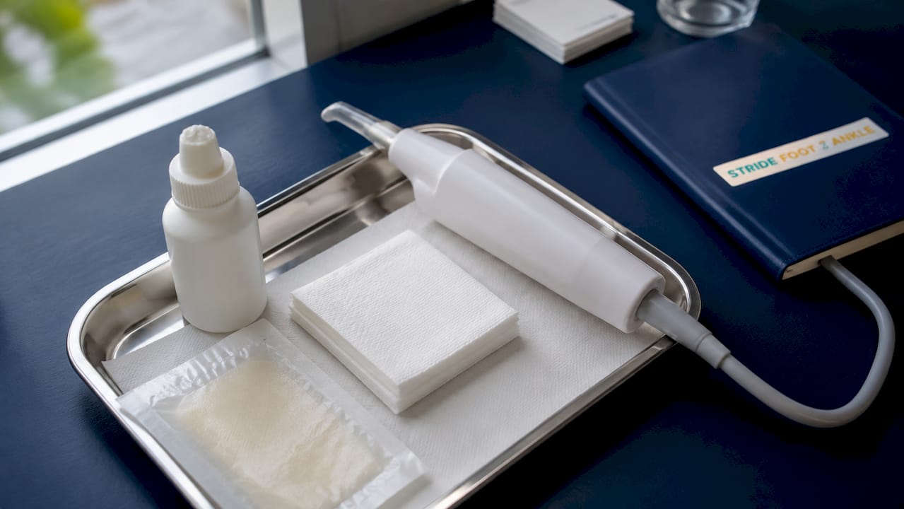
What are the different types of foot wound debridement?
No single method works for every wound. The choice depends on wound depth, infection status, the patient’s circulation, and pain tolerance. A 2026 systematic review confirmed that method selection should always reflect an individualized assessment rather than a one-size approach.
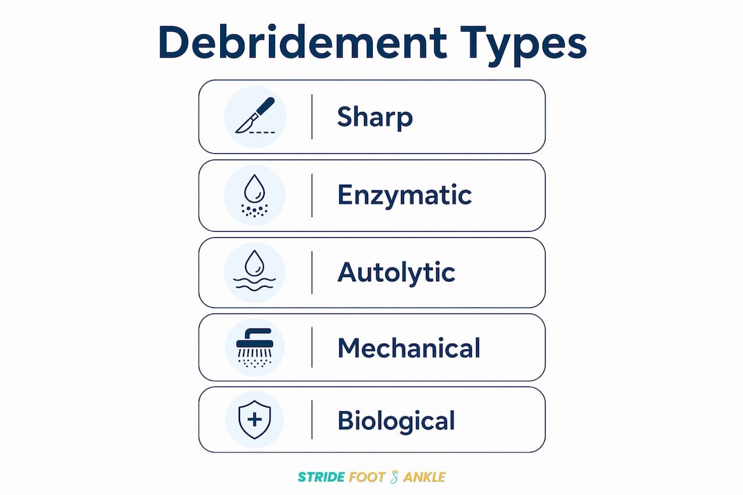
Sharp surgical debridement
Sharp debridement uses scalpels, curettes, or scissors to cut away necrotic tissue directly. It is the first-line method for most neuropathic foot ulcers and infected wounds because it works immediately and allows the clinician to see and control exactly what is removed. It requires a trained clinician and is typically performed in an office or clinical setting.
Enzymatic debridement
Enzymatic debridement applies a prescription topical agent, such as collagenase, directly to the wound. The enzyme breaks down dead tissue chemically over several days. This method suits patients who cannot tolerate sharp debridement due to poor circulation or bleeding risk. It is slower but gentler, and it works well as a bridge between sharp sessions.
Autolytic debridement
Autolytic debridement uses the body’s own moisture and enzymes, kept in place by a moisture-retentive dressing, to soften and dissolve necrotic tissue. It is the least aggressive option and works best for shallow wounds with minimal infection. The process takes days to weeks and is not appropriate when infection is present or advancing.
Mechanical debridement
Mechanical methods include wet-to-dry dressings, wound irrigation, and ultrasound-assisted debridement. Ultrasound is particularly useful for disrupting biofilms that are invisible to the naked eye. Effective debridement often requires multi-modal approaches that combine sharp techniques with ultrasound to reach bacteria embedded deep in the wound bed.
Biological debridement
Biological debridement uses medical-grade maggots placed on the wound. The larvae consume only necrotic tissue and secrete enzymes that promote healing. A 2026 systematic review found that biological debridement showed a higher healing likelihood compared to standard care in some studies, with a relative risk of 4.51. This method is typically reserved for complex wounds that have not responded to other approaches.
| Method | How it works | Best for | Limitations |
|---|---|---|---|
| Sharp surgical | Scalpel or curette removes tissue directly | Infected ulcers, neuropathic wounds | Requires trained clinician, may need anesthesia |
| Enzymatic | Topical agent dissolves dead tissue | Patients with bleeding risk or poor circulation | Slow, prescription required |
| Autolytic | Moisture-retentive dressing activates body enzymes | Shallow, minimally infected wounds | Too slow for infected or deep wounds |
| Mechanical | Irrigation or ultrasound disrupts tissue and biofilm | Biofilm-heavy wounds | Can damage healthy tissue if used incorrectly |
| Biological | Medical maggots consume necrotic tissue | Complex, non-healing wounds | Limited availability, patient acceptance varies |
Key factors that guide method selection:
- Wound depth and size: Deeper wounds with tunneling require sharp or mechanical methods.
- Infection level: Active infection demands sharp debridement for speed.
- Circulation status: Poor blood flow rules out aggressive sharp techniques until vascular status is assessed.
- Patient health: Anticoagulant use, diabetes severity, and immune status all affect which method is safe.
How does foot wound debridement promote healing and reduce complications?
Debridement works on two levels at once: it removes what is blocking healing and it actively stimulates the body to restart repair. Dead tissue does not just sit passively in a wound. It releases inflammatory signals that keep the wound locked in a chronic state and feeds bacterial colonies that compete with healing cells for nutrients and oxygen.
Debridement clears infection, removes physical and biochemical barriers, and stimulates blood flow and growth factors. That combination transforms a stalled wound into one that progresses through the normal healing stages. Without it, even the best dressings and antibiotics cannot overcome the underlying blockage.
The benefit is especially significant for diabetic foot ulcers. Diabetes impairs nerve function and circulation, which means wounds often go unnoticed until they are deep and infected. The tissue in these wounds contains high numbers of senescent cells, old cells that no longer divide but release damaging enzymes. Removing senescent cells through debridement shifts the wound environment from destructive to regenerative.
“Debridement reduces bioburden and removes senescent cells that prevent wounds from healing, shifting chronic wounds into an active healing state. This shift is especially critical for diabetic foot ulcers, where the risk of amputation rises sharply when wounds stall without intervention.”
Preventing amputation is the highest-stakes reason to take debridement seriously. Chronic foot ulcers that do not receive timely and appropriate wound treatment are a leading pathway to lower-limb amputation in patients with diabetes. Consistent debridement, combined with foot ulcer prevention strategies, significantly reduces that risk by keeping wounds on an active healing trajectory.
Vascular assessment before aggressive debridement is also critical. Poor circulation can worsen a wound by creating larger non-healing injuries if blood flow is insufficient to support tissue repair after removal. A thorough vascular evaluation protects patients from procedures that could do more harm than good.
What should patients know about recovery after foot wound debridement?
Recovery after debridement is generally manageable, but knowing what is normal versus what requires a call to your clinician makes a real difference in outcomes. Mild soreness at the wound site is expected for 1–2 days after the procedure. Over-the-counter pain medications like acetaminophen or ibuprofen handle this well for most patients.
Watch for these warning signs that require immediate clinical contact:
- Fever above 101°F: This signals a possible systemic infection spreading beyond the wound.
- Thick green or yellow pus: Purulent discharge indicates active bacterial infection in the wound bed.
- Foul odor from the wound: A new or worsening odor suggests bacterial overgrowth or tissue breakdown.
- Spreading redness around the wound: Redness that extends beyond the wound edges points to cellulitis, a skin infection that spreads quickly.
- Increased swelling or warmth: These signs suggest the wound is not responding well and needs reassessment.
Normal recovery includes mild soreness managed with over-the-counter medications, with clear instructions on warning signs like fever, foul odor, and increased redness. If you experience any of the warning signs above, contact your clinician the same day rather than waiting for your next scheduled appointment.
Chronic wounds typically require repeated debridement sessions every 1–2 weeks. This frequency is not a sign that treatment is failing. It reflects the reality that necrotic tissue can reform between sessions, especially in wounds with poor circulation or ongoing infection. Consistent follow-up is the most reliable path to closure.
Mobility guidance varies by wound location and depth. Your clinician will advise on weight-bearing restrictions. For plantar wounds on the bottom of the foot, offloading with a specialized boot or cast is often necessary to prevent pressure from reopening the wound bed between sessions. Detailed post-procedure care steps help you manage recovery safely at home.
Pro Tip: Keep a simple wound log between appointments. Note any changes in color, odor, drainage, or pain level. This record gives your clinician precise information and speeds up decision-making at each visit.
Key Takeaways
Foot wound debridement is the single most important step in restarting a stalled wound’s healing process, and no dressing or antibiotic replaces it.
| Point | Details |
|---|---|
| Core definition | Debridement removes necrotic, infected, or damaged tissue to restart the healing cascade. |
| Method selection | Sharp surgical debridement is first-line; enzymatic, autolytic, mechanical, and biological methods suit specific wound types. |
| Healing mechanism | Debridement clears bioburden, removes senescent cells, and stimulates growth factors to shift chronic wounds to active healing. |
| Recovery expectations | Mild soreness lasts 1–2 days; fever above 101°F, pus, or spreading redness requires same-day clinical contact. |
| Repeat sessions | Chronic wounds need debridement every 1–2 weeks; consistent follow-up is the most reliable path to wound closure. |
What I have learned from watching patients go through debridement
The biggest misconception patients bring into a debridement appointment is that the procedure is purely aggressive, a kind of medical scraping that causes more damage than it fixes. That fear is understandable, but it misrepresents what skilled clinicians actually do. Debridement is a sculpting process. The goal is always to remove exactly what is blocking healing while leaving every viable cell intact.
What I have seen change outcomes most is not the method chosen but the conversation before and during the procedure. Patients who understand why each step happens tolerate the process better and communicate more clearly when something feels wrong. That communication is not a distraction. It is clinical data. Sharp pain during debridement tells the clinician to stop and reassess, which protects healthy tissue.
The vascular assessment piece is the one most often skipped in rushed clinical settings, and it is the one that matters most for safety. Aggressive debridement on an ischemic foot, one with severely restricted blood flow, can create a wound larger than the one you started with. A thorough circulation check before any procedure is not optional. It is the foundation of responsible wound care.
Patients with diabetic foot ulcers sometimes feel discouraged when they need multiple sessions. The honest truth is that repeated debridement is a sign the treatment plan is working, not failing. Each session removes another layer of the problem. Staying consistent with follow-up appointments, combined with comprehensive foot care, is what gets wounds across the finish line.
— Ramil
Wound care expertise at Stridefootankle in Las Vegas
Stridefootankle provides specialized wound care services under the direction of Dr. Nahad Wassel, a board-certified podiatric surgeon with advanced training in foot and ankle surgery. Every patient receives a thorough evaluation that includes vascular assessment, wound staging, and a personalized treatment plan before any procedure begins.

Whether you are managing a diabetic foot ulcer, a post-surgical wound, or a chronic injury that has not responded to basic care, Stridefootankle offers the clinical depth to move your healing forward. Dr. Wassel’s patient-centered approach means you receive clear explanations at every step and a care plan built around your specific wound and health status. Schedule a consultation through the wound care services page to get a professional evaluation and start striding confidently toward recovery.
FAQ
What is foot wound debridement in simple terms?
Foot wound debridement is the medical removal of dead, infected, or damaged tissue from a foot wound. This clears the way for healthy tissue to grow and reduces the risk of serious infection.
Does foot wound debridement hurt?
Most patients feel pressure or pulling during the procedure, not sharp pain, because local anesthesia is used. Sharp pain signals contact with healthy tissue and should be reported to the clinician immediately so technique can be adjusted.
How often do chronic wounds need debridement?
Chronic wounds typically require debridement every 1–2 weeks. This frequency reflects the ongoing nature of tissue breakdown in stalled wounds, not a failure of treatment.
Which type of debridement is best for diabetic foot ulcers?
Sharp surgical debridement is the first-line method for most diabetic foot ulcers because it works immediately and removes infected tissue precisely. Enzymatic or autolytic methods may be added as adjuncts based on wound characteristics and the patient’s circulation.
When should I call my clinician after a debridement procedure?
Contact your clinician the same day if you develop a fever above 101°F, notice thick pus or a foul odor, or see redness spreading beyond the wound edges. Mild soreness for 1–2 days after the procedure is normal and manageable with over-the-counter pain medication.
Recommended
- Wound Care – Stride Foot & Ankle – Dr. Nahad Wassel
- Wounds: Types, Healing Stages, and Treatment Guide – Stride Foot & Ankle – Dr. Nahad Wassel
- Step-by-step foot care guide for Las Vegas residents – Stride Foot & Ankle – Dr. Nahad Wassel
- Post-Surgery Foot Care Steps for Safe Recovery – Stride Foot & Ankle – Dr. Nahad Wassel
Recent Comments