

TL;DR:
- Imaging in podiatry extends beyond X-rays, with MRI, ultrasound, and nuclear scans guiding diagnosis and treatment. Proper modality selection and timing are crucial for early detection of conditions like Charcot foot and diabetic osteomyelitis, improving patient outcomes. Integrating advanced imaging with clinical judgment prevents misdiagnosis and optimizes care.
The role of imaging in podiatry goes far beyond a quick X-ray. Accurate diagnosis of foot and ankle conditions depends on matching the right imaging tool to the right clinical question. From detecting early Charcot neuropathic osteoarthropathy before bone collapse sets in, to monitoring diabetic foot ulcer healing remotely, modern podiatric imaging directly shapes treatment decisions and patient outcomes. This guide breaks down every major modality, its real-world applications, and the clinical reasoning that ties them together.
Table of Contents
- Key Takeaways
- Core imaging modalities in podiatric practice
- Advanced imaging for Charcot foot and diabetic infections
- Ultrasound and digital imaging in wound and soft tissue care
- Clinical decision-making: building an imaging strategy
- Pitfalls and future directions in podiatric imaging
- My perspective on getting imaging right
- How Stridefootankle uses advanced imaging for your care
- FAQ
Key Takeaways
| Point | Details |
|---|---|
| X-rays are a starting point | Plain radiographs are first-line but often normal in early disease, requiring escalation to MRI or advanced imaging. |
| MRI leads in soft tissue diagnosis | MRI detects early bone marrow edema and distinguishes Charcot foot from osteomyelitis when X-rays appear normal. |
| Ultrasound offers dynamic evaluation | High-resolution ultrasound identifies ligament injuries, tendon pathology, and muscle atrophy without radiation exposure. |
| Nuclear imaging fills diagnostic gaps | WBC SPECT/CT outperforms MRI at initial diabetic foot osteomyelitis diagnosis and clarifies ambiguous post-treatment scans. |
| Digital wound imaging supports remote care | Digital imaging of diabetic foot ulcers aids documentation and remote consultation, with machine learning improving assessment accuracy. |
Core imaging modalities in podiatric practice
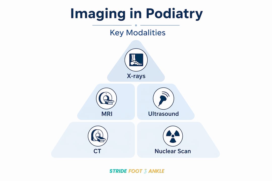
The right imaging modality depends on what you are looking for. Plain radiographs, MRI, ultrasound, CT, and nuclear medicine each reveal a different layer of foot and ankle pathology. Understanding when to use each one is the foundation of good diagnostic imaging in podiatric practice.
Plain radiographs
Plain radiographs remain the first step in evaluating most foot and ankle complaints. They are fast, widely available, and cost-effective for identifying fractures, joint space narrowing, deformity, and calcification. A standard three-view ankle series covers the majority of bony concerns. That said, radiographs are often normal in early stages of conditions like Charcot neuropathic osteoarthropathy, which means a normal X-ray does not rule out serious pathology.
MRI
MRI is the gold standard for soft tissue evaluation and early detection of bone marrow changes. It excels at identifying stress fractures, ligament tears, tendon pathology, occult fractures, and the bone marrow edema that signals early Charcot disease. When symptoms persist despite normal radiographs, MRI is recommended as the next step. For foot and ankle care involving complex or persistent pain, MRI provides a level of tissue detail no other modality matches.
Ultrasound
Ultrasound is radiation-free, portable, and uniquely capable of real-time, dynamic assessment. A provider can compress a tendon during the scan, observe movement, or guide an injection precisely. High-resolution ultrasound is gaining traction as a first-line soft tissue imaging tool given its accessibility and safety profile.
CT and nuclear imaging
CT scanning provides exceptional bony detail for surgical planning, hardware evaluation, and complex deformity assessment. When infection status is uncertain or MRI is contraindicated, nuclear imaging modalities like white blood cell (WBC) SPECT/CT step in. They are particularly valuable when distinguishing active infection from post-surgical changes or inflammatory arthropathy.
Pro Tip: When ordering imaging for foot and ankle pain, always pair the imaging choice with a specific clinical question. “Rule out pathology” is not a useful driver. “Is this bone marrow edema from infection or Charcot?” is.
Advanced imaging for Charcot foot and diabetic infections
Advanced imaging is most critical when the clinical stakes are highest. Two conditions where getting the diagnosis wrong carries severe consequences are Charcot neuropathic osteoarthropathy and diabetic foot osteomyelitis. Both can look similar on exam and both can be missed on plain radiographs.
Charcot foot detection
Charcot neuropathic osteoarthropathy is a progressive joint destruction process that occurs in patients with peripheral neuropathy, most commonly from diabetes. In the acute phase, the foot appears red, swollen, and warm, which is frequently mistaken for cellulitis or deep vein thrombosis. The critical clinical trap is that early Charcot radiographs can appear completely normal. By the time bony fragmentation appears on X-ray, significant joint destruction has already occurred.
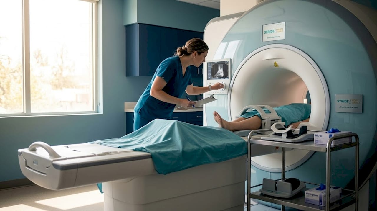
MRI changes that picture entirely. It reveals bone marrow edema, subchondral cysts, joint effusion, and periarticular soft tissue inflammation in the acute stage, before any radiographic abnormality is visible. When your clinical suspicion is high and the X-ray looks normal, MRI should be prioritized without delay. Missing early Charcot leads to progressive collapse, deformity, and ultimately amputation risk.
Differentiating Charcot from osteomyelitis
Distinguishing Charcot from osteomyelitis is one of the most challenging problems in diabetic foot imaging. Both produce bone marrow edema on MRI. Both cause bony destruction over time. The treatment paths, however, are completely different. Osteomyelitis requires antibiotics or surgical debridement. Charcot requires offloading and protected weight bearing.
MRI is first-line for suspected diabetic foot complications, but when MRI findings are equivocal or the patient cannot undergo MRI, nuclear imaging becomes the deciding tool. A 2026 prospective study found that WBC SPECT/CT exceeded MRI in sensitivity, specificity, positive predictive value, negative predictive value, and ROC area at initial diagnosis of diabetic foot osteomyelitis. After six to eight weeks of antibiotic therapy, both modalities performed similarly for monitoring treatment response.
| Imaging modality | Initial osteomyelitis diagnosis | Post-treatment monitoring |
|---|---|---|
| MRI | Strong, first-line choice | Good, but confounders apply |
| WBC SPECT/CT | Superior diagnostic performance | Comparable to MRI |
| Plain radiograph | Insensitive in early disease | Limited utility |
| CT | Bony detail only | Useful for structural assessment |
Pro Tip: If a patient with diabetes has bone marrow edema on MRI after completing antibiotic therapy, do not assume the infection has resolved. Prior surgery, trauma, and Charcot changes can all produce persistent edema. WBC SPECT/CT can clarify the picture in these ambiguous cases.
Ultrasound and digital imaging in wound and soft tissue care
Ultrasound and digital imaging tools address a different but equally important clinical need. They make diagnosis faster, more accessible, and in many cases, less expensive. Their role in the role of imaging in foot wounds and soft tissue evaluation is growing steadily.
High-resolution ultrasound for soft tissue pathology
A study of 40 patients demonstrated that high-resolution ultrasound (HRUS) identified ligament injuries in 36% of cases, tendon pathology in 28%, and plantar fasciitis in 8%, along with other soft tissue findings across the group. These are real-world numbers from a clinical setting, and they reflect how much soft tissue detail HRUS can capture without any ionizing radiation.
The dynamic capability of ultrasound sets it apart. A clinician can ask the patient to dorsiflex the ankle and watch the Achilles tendon in real time. That kind of functional information is simply unavailable on a static MRI. For suspected plantar fasciitis, Achilles tendinopathy, or peroneal subluxation, ultrasound is often the most direct and informative next step after clinical exam.
For patients with diabetes, the benefits extend further. Ultrasound assesses intrinsic foot muscle atrophy early and non-invasively, detecting changes in muscle bulk and real-time contraction that may precede clinical signs of neuropathy-related weakness. It is more accessible than MRI, requires no contrast, and produces no radiation exposure. For patients who need regular monitoring, that matters.
Digital wound imaging and its limitations
Digital wound imaging gives clinicians a way to document, measure, and share diabetic foot ulcer data across care teams. It supports monitoring of wound dimensions, tissue characteristics, and healing trajectory over time. When combined with telehealth platforms, it enables remote wound consultation with specialists, which is particularly relevant for patients in Las Vegas and surrounding areas who may have limited access to subspecialty care.
The limitation is real and worth knowing. Remote photo assessments of diabetic foot ulcers show low-to-moderate reliability for infection signs like exudate and periwound changes. What looks like simple maceration in a photo may be early infection in person. The solution being developed involves machine learning models trained on standardized wound photo protocols, which have the potential to improve remote assessment accuracy and support telemedicine care models significantly. Stridefootankle follows advances in this area closely, particularly as it relates to diabetic foot ulcer prevention and wound care management.
Clinical decision-making: building an imaging strategy
The role of imaging in foot diagnosis is not just about knowing what each modality can do. It is about knowing when to use each one, in what sequence, and how to integrate those findings with what you see and hear from the patient. A systematic approach prevents both under-imaging and unnecessary testing.
Here is a practical framework for clinical imaging decisions in podiatry:
Start with plain radiographs. For almost every new foot or ankle complaint, three-view radiographs are the first step. They rule out fractures, identify alignment issues, and detect calcification or degenerative change quickly and affordably.
Escalate to MRI when symptoms persist. If a patient has had persistent pain for four to six weeks despite conservative care and normal radiographs, MRI is the next logical step. It picks up stress fractures, early marrow edema, and soft tissue injuries that X-rays miss entirely.
Choose ultrasound for targeted soft tissue questions. Suspected tendinopathy, ligament sprain, plantar fasciitis, or the need for injection guidance all point to ultrasound as the right tool. It is faster to schedule, involves no radiation, and provides dynamic information.
Use bilateral imaging for diagnosis, not just the symptomatic side. Bilateral foot radiographs help detect subtle asymmetry in Charcot, flatfoot deformity, and alignment changes that would be missed on a unilateral study alone.
Bring in nuclear imaging for complex or equivocal cases. When the diagnosis is uncertain after MRI, when MRI is contraindicated, or when you need to distinguish active infection from post-treatment edema, WBC SPECT/CT adds a critical layer of specificity.
Use CT for surgical planning. When a patient is heading toward reconstructive surgery or you need precise anatomical mapping of a complex fracture or deformity, CT provides the bony architecture detail that guides safe, effective intervention.
Pro Tip: Correlation between imaging findings and the clinical exam is not optional. Bone marrow edema on MRI means nothing in isolation. A red, swollen, warm foot in a diabetic patient with peripheral neuropathy plus marrow edema on MRI is an entirely different conversation. Always read imaging in clinical context.
For a detailed walkthrough of how these decisions play out in practice, Stridefootankle’s foot pain diagnosis guide covers the full diagnostic pathway from first visit to treatment planning.
Pitfalls and future directions in podiatric imaging
Every imaging modality has blind spots. Recognizing them is as important as knowing what each modality can find. The importance of imaging in foot health depends just as much on avoiding misinterpretation as it does on ordering the right test.
The most consequential pitfall in podiatric imaging is the false reassurance of a normal X-ray:
- Early Charcot with normal radiographs: As noted, radiographs can be deceptively normal in acute Charcot. A patient with diabetes, peripheral neuropathy, and a hot swollen foot needs MRI even when the X-ray shows nothing. Waiting for radiographic changes means waiting for bone collapse.
- Post-surgical MRI interpretation: Bone marrow edema persists after surgery, trauma, and in the setting of Charcot changes. MRI specificity for infection resolution drops significantly in patients with prior foot surgery. Misreading residual edema as ongoing infection leads to unnecessary antibiotics or repeat procedures.
- Ultrasound operator dependence: Ultrasound quality depends heavily on the skill of the operator. A poorly performed study can miss a partial tendon tear or mischaracterize a ligament injury. Standardized training and experience are non-negotiable for reliable results.
- Digital wound photo limitations: Remote infection assessment via photos remains unreliable without AI-assisted interpretation. Using digital wound images as the sole basis for antibiotic decisions is a clinical risk without validated decision-support tools in place.
Looking forward, several technologies are reshaping diagnostic imaging in podiatric practice. Dual-energy CT can differentiate gout deposits from calcium pyrophosphate without biopsy, which has direct relevance for arthropathy workup in the foot. FDG-PET/CT is emerging as a complementary tool for complex infection and tumor workup. Machine learning models trained on wound photography show real promise for improving the accuracy of remote diabetic foot assessment.
“The future of podiatric imaging is not one modality replacing another. It is a smarter integration of each tool’s strengths, guided by clinical reasoning and patient-specific context.”
The clinicians and patients who will benefit most from these advances are those who understand that imaging informs judgment. It does not replace it.
My perspective on getting imaging right
I have seen what happens when imaging decisions are made reflexively rather than thoughtfully. A patient with a hot, red foot gets an X-ray, the X-ray looks normal, and the team sends them home with a cellulitis diagnosis. Three weeks later, they return with Charcot collapse that is now surgical. The X-ray was not wrong. It just was not enough.
What I have found in practice is that the most common imaging mistake is not ordering the wrong test. It is stopping too early. X-rays are a starting point, not an endpoint. The moment a result does not match the clinical picture, that is your signal to go further.
I have also learned that cost and access matter in real patient care. Not everyone can get an MRI quickly. In those situations, I lean on high-resolution ultrasound more than many clinicians expect. It is underutilized for early tendon and ligament pathology, and it provides immediate, dynamic information in the room. Used well, it reduces the number of MRI referrals for straightforward soft tissue questions.
My honest advice to both clinicians and patients: ask what the imaging is supposed to answer before ordering it. If the answer is vague, the imaging will be vague too. The best outcomes come from specific clinical questions matched to the right modality. That is the part that no machine, no matter how advanced, can do for you.
— Ramil
How Stridefootankle uses advanced imaging for your care

At Stridefootankle, Dr. Nahad Wassel integrates the full spectrum of podiatric imaging into every patient’s diagnostic and treatment plan. Whether you need plain radiographs for a suspected fracture, advanced imaging for a complex diabetic foot concern, or specialized ultrasound wound healing therapy for a non-healing ulcer, the practice brings the right tools to your specific situation.
Las Vegas patients have access to personalized, imaging-guided care that does not stop at a single test. Dr. Wassel combines clinical expertise with precise diagnostics to ensure your treatment plan reflects what is actually happening in your foot and ankle. If you have been dealing with persistent foot pain, a wound that will not heal, or a diagnosis that has not felt quite right, the foot and ankle care services at Stridefootankle are a strong next step. Request an appointment today and get clarity, not just a scan.
FAQ
What imaging is used first for foot and ankle pain?
Plain radiographs (X-rays) are the standard first-line imaging for most foot and ankle complaints. They assess bone structure, alignment, and deformity quickly. If symptoms persist despite normal X-rays, MRI is typically the next step.
When is MRI necessary in podiatry?
MRI is recommended when plain radiographs are normal but symptoms persist, when soft tissue injury is suspected, or when early Charcot neuropathic osteoarthropathy or osteomyelitis needs to be ruled out. It offers unmatched detail for bone marrow changes and soft tissue pathology.
Is ultrasound useful in podiatry?
Yes. High-resolution ultrasound identifies ligament injuries, tendon abnormalities, plantar fasciitis, and muscle atrophy without radiation. Its dynamic imaging capability makes it especially useful for tendon and soft tissue assessment in real time.
How is imaging used in diabetic foot care?
In diabetic foot care, imaging guides the detection of osteomyelitis, Charcot neuropathic osteoarthropathy, and wound depth. MRI is first-line for bone and soft tissue evaluation, while WBC SPECT/CT is used when MRI findings are ambiguous or post-treatment monitoring is needed.
Can digital photos replace clinical wound assessment?
Digital wound images support documentation and remote consultation but show low-to-moderate reliability for detecting infection signs remotely. They work best as a supplement to in-person evaluation, not a replacement, particularly for diabetic foot ulcers where infection can be subtle.
Recommended
- Why See a Podiatrist? Your Complete Foot Health Guide – Stride Foot & Ankle – Dr. Nahad Wassel
- Foot pain podiatry: Las Vegas diagnosis and treatment guide – Stride Foot & Ankle – Dr. Nahad Wassel
- How Patient-Centered Care Transforms Podiatry in Las Vegas – Stride Foot & Ankle – Dr. Nahad Wassel
- General Foot & Ankle Care – Stride Foot & Ankle
Recent Comments