

Most stable, nondisplaced Lisfranc injuries heal with strict non-weightbearing immobilization for 6 to 8 weeks without a scalpel involved. Unstable, displaced, or comminuted injuries usually need surgical fixation, whether that’s screws, plates, or joint fusion, to restore alignment and prevent long-term arthritis. Recovery ranges from a few weeks for a mild sprain treated conservatively to several months after surgery for a severe fracture-dislocation.
TL;DR:
- Conservative treatment is suitable only when initial weight-bearing X-rays show no displacement and the gap between the first and second metatarsals stays under 3 mm.
- Imaging, especially at two weeks post-injury, is critical to detect any secondary displacement that may necessitate surgery.
- Surgery options like screw or plate fixation, bridge plating, or fusion depend on injury severity, with displaced or unstable injuries almost always requiring operative intervention.
- Post-surgery recovery typically involves six weeks of non-weightbearing followed by progressive weightbearing, physical therapy, and sport-specific rehab over several months.
- Long-term outcomes rely heavily on precise bone placement during treatment, with residual displacement over 2 mm linked to increased arthritis and chronic foot problems.
Table of Contents
- Lisfranc Injury Treatment: An Overview of Your Options
- What Conservative Lisfranc Injury Care Actually Involves
- How Surgeons Treat Unstable Lisfranc Injuries
- What Determines Whether You Need Surgery?
- The Lisfranc Recovery Timeline: What to Expect
- Complications and Long-Term Outlook
- Why Choose Stride Foot & Ankle for Lisfranc Injury Care
- An Editorial Take on Treating This Injury
- Ready for a Lisfranc Evaluation? Here’s How to Book One
- Sources
- FAQ
Lisfranc Injury Treatment: An Overview of Your Options
Lisfranc injury treatment splits into two paths, and the fork in the road is stability. If your midfoot joints stay aligned when you bear weight, your foot has a good shot at healing in a boot or cast. If the bones have shifted, or shift under load, surgery becomes the more reliable route to a functional foot.
Conservative care exists to protect a joint that is already in the right position. It’s not a lesser option; it’s the correct option when the anatomy supports it. Surgery exists to put bones back where they belong and hold them there long enough for ligaments and joints to stabilize.
Surgeons choose among a handful of established techniques, each with a different tradeoff between rigidity and joint preservation:
- Open reduction internal fixation (ORIF) with screws or plates, the most common approach for displaced fractures.
- Bridge plating, which spans the joint without drilling through cartilage.
- Primary arthrodesis (fusion), fusing the affected joints permanently, often used for purely ligamentous or severely comminuted injuries.
- Suture-button fixation, a flexible, joint-sparing option gaining traction for select ligament injuries, especially in athletes.
Nonoperative management, meanwhile, follows a fairly predictable script: a period of complete non-weightbearing, imaging to confirm nothing has shifted, then a gradual return to walking. The details of that protocol matter more than most patients realize, which is why the next section walks through it step by step.
What Conservative Lisfranc Injury Care Actually Involves
Conservative treatment for lisfranc sprain and stable fracture patterns centers on one non-negotiable rule: keep weight off the foot for 6 to 8 weeks while the ligaments and bones reunite. This works only when weight-bearing X-rays show the joint holding its position and the diastasis between the first and second metatarsals stays small.
You’re a candidate for conservative care if your injury meets a few specific criteria. Weight-bearing radiographs need to show no meaningful displacement. The gap between the base of the first and second metatarsals, what surgeons call the M1 to M2 distance, needs to stay under the threshold that typically pushes cases toward surgery. And the injury pattern needs to look like a soft-tissue sprain rather than a fracture-dislocation.
Here’s what the actual protocol looks like once you’re cleared for nonoperative management:
- Strict non-weightbearing immobilization for 6 to 8 weeks, supported by clinical evidence on Lisfranc management, in a cast or a removable boot, depending on your surgeon’s preference and how well you can be trusted to actually stay off it.
- Repeat weight-bearing X-rays around the two-week mark, the point where secondary displacement, if it’s going to happen, tends to reveal itself.
- Transition to a protected weight-bearing boot once imaging confirms the joint has held, usually somewhere between weeks 6 and 8.
- Gradual return to a supportive shoe with continued monitoring for pain, swelling, or any sense of instability.
The risk with conservative care isn’t that it fails outright. It’s that a joint that looked stable on day one shifts slightly over the following weeks, which is exactly why the two-week follow-up film isn’t optional. If that image shows new displacement, the conversation about surgery starts over.
Pro Tip: Elevate your foot above heart level for the first two weeks, even in the boot. Swelling that lingers doesn’t just hurt, it can mask early signs of shifting alignment on your follow-up X-ray.
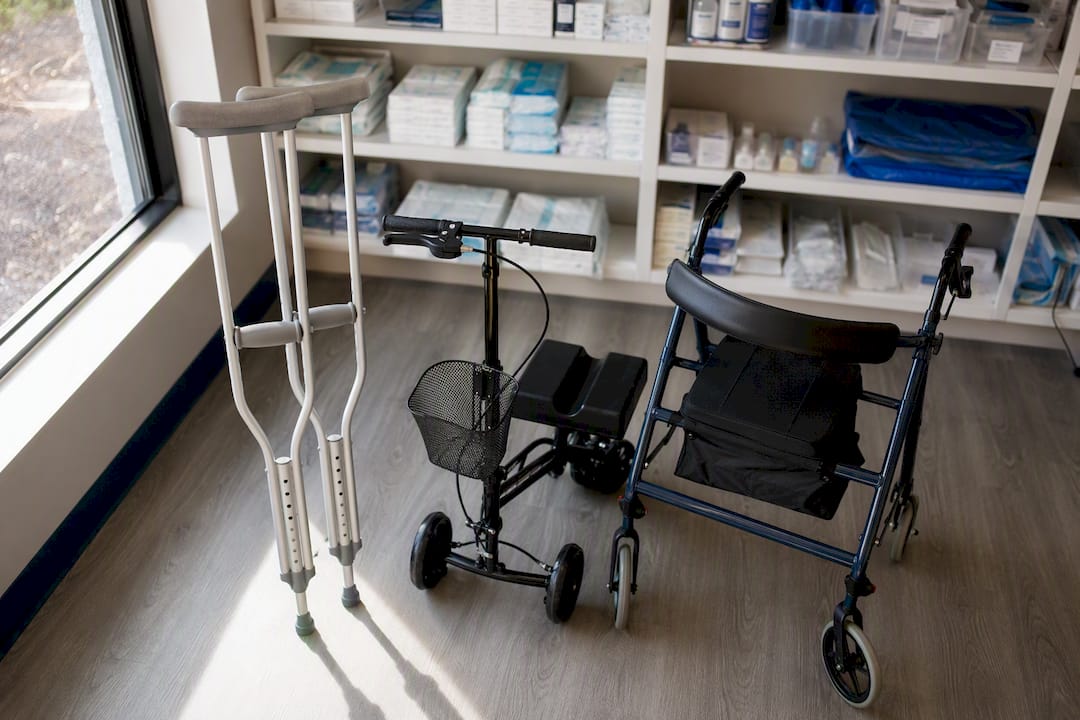
Crutches, a knee scooter, or a wheeled walker all work for staying non-weightbearing; which one you choose usually comes down to your home layout and upper body strength. Our guide to nonsurgical options for tendon and ligament injuries covers boot and cast choices in more depth if you want to compare them before your next appointment.
How Surgeons Treat Unstable Lisfranc Injuries
Surgery becomes the recommended path once imaging shows true instability, meaningful displacement, or a fracture pattern too comminuted to trust in a boot. The goal is anatomic reduction: getting every bone back to its precise original position, because how close that reduction comes to perfect is the single biggest factor in how your foot functions years later.
Surgeons generally reach for one of a few techniques, and the choice depends on the injury pattern rather than personal preference alone.
- ORIF with screws or plates remains the workhorse for displaced fracture-dislocations, giving rigid fixation while the joint heals.
- Bridge plating is increasingly favored over screws that cross the joint directly. Some surgical teams prefer dorsal bridge plating because it achieves comparable reduction while sparing cartilage, which matters if you’re hoping to avoid a fused joint down the road.
- Primary arthrodesis fuses the joint outright and tends to be the pick for purely ligamentous injuries or fracture patterns too shattered to reconstruct. Evidence on Lisfranc dislocation suggests fusion can produce better patient-reported outcomes and fewer reoperations than ORIF in select cases.
- Suture-button fixation offers a flexible alternative for ligament-dominant injuries, particularly in athletes who want to avoid permanent hardware crossing the joint.
Hardware choice carries its own tradeoffs. Screws and plates that cross the joint directly tend to irritate surrounding tissue, and a fair number of patients end up back in the operating room later just to have that hardware removed. Joint-sparing alternatives like bridge plating and suture-button constructs appear to lower that secondary-procedure risk, since nothing rigid is left grinding across the joint surface long term.
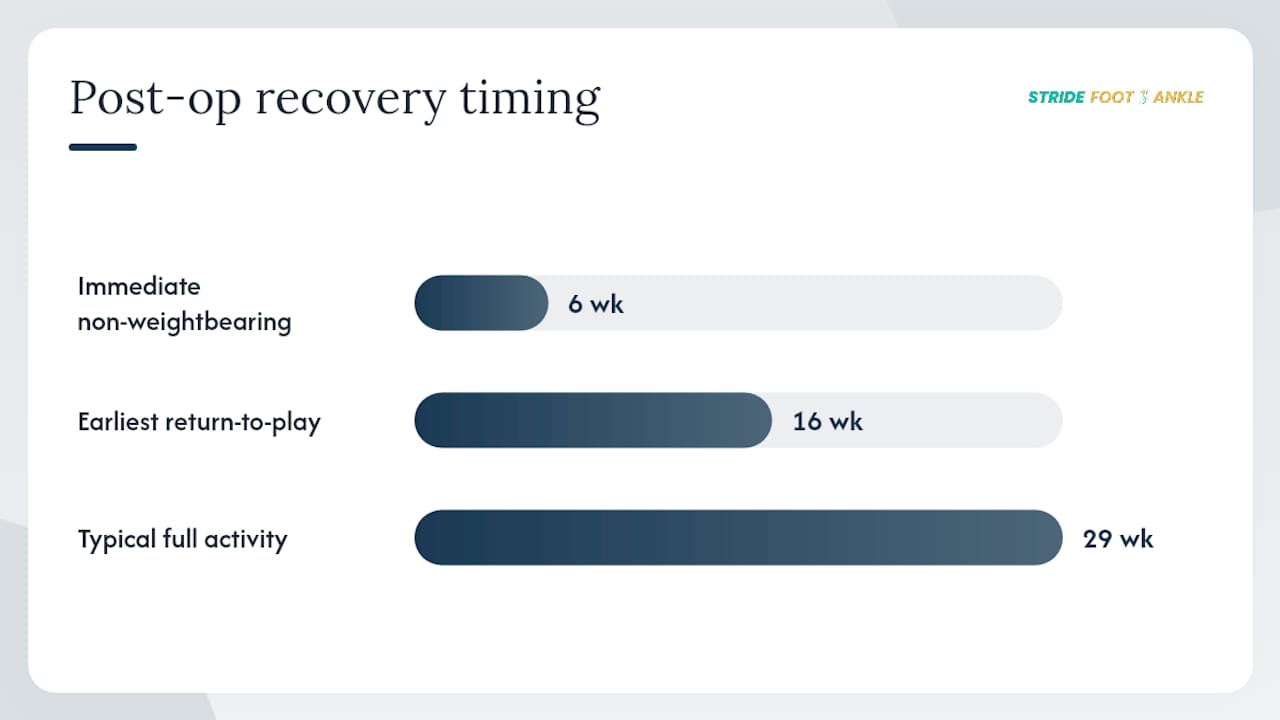
By the numbers: Return-to-play rates after surgical treatment for Lisfranc injuries range from 86% to 100%, with the average athlete cleared for full activity somewhere between 16 and 29 weeks after surgery, depending on injury severity and fixation type.
Immediately after surgery, expect a period of non-weightbearing similar to the conservative pathway, typically around six weeks in a cast or boot, with close attention to the incision for early signs of infection or wound breakdown. Our post-surgery foot care guide walks through wound monitoring and hardware care in more detail if you’re heading into an operation.
What Determines Whether You Need Surgery?
The decision between conservative care and surgery rests on imaging, not guesswork. Surgeons weigh weight-bearing X-rays first, add CT or MRI when the picture is unclear, and compare the result against established diastasis thresholds and displacement patterns before recommending a path.
Weight-bearing radiographs come first because a Lisfranc injury can look stable on a resting film and reveal instability the moment you put weight on it. That’s the trap that catches missed diagnoses. If pain and exam findings suggest midfoot instability but standard films look clean, maintaining a high index of suspicion and ordering advanced imaging is what catches injuries that would otherwise slip through.
CT scans map bony fragments and subtle displacement that plain film misses, while MRI is more useful for assessing ligament integrity when the bones themselves look intact but the joint still feels unstable on exam.
The specific numbers surgeons use include:
- Diastasis of roughly 2 to 3 mm between the first and second metatarsal bases; one cohort study found a cutoff near 3.0 mm was associated with a shift toward operative treatment.
- Buehren qualitative signs (A/C), a classification that looks at displacement patterns rather than a single measurement.
- Talo-metatarsal angulation, flagging rotational or angular displacement that a diastasis measurement alone can miss.
- Myerson and Hardcastle classification systems, which categorize injury patterns by direction and severity of displacement to guide treatment planning.
No single number decides your treatment. A diastasis measurement near the cutoff combined with qualitative displacement signs and a high-energy trauma mechanism, a car accident versus a rolled ankle playing pickup basketball, tips the decision in different directions even at similar measurements. That two-week follow-up film after conservative care matters for the same reason: subtle instability sometimes only reveals itself once weight-bearing starts. If your surgeon and you disagree on which side of that line your injury falls, a second opinion reviewing the same films is a reasonable ask, not an insult. Our imaging guide breaks down what each scan type is actually looking for.
The Lisfranc Recovery Timeline: What to Expect
Recovery follows a staged progression from strict non-weightbearing to full activity, and rushing any stage is the most common way patients set themselves back. Most people return to normal activity somewhere between 16 and 29 weeks after surgery, while conservative cases often finish sooner if the joint stays stable throughout.
Whether you had surgery or a boot, the early weeks look similar: complete non-weightbearing, elevation, and controlling swelling before it becomes chronic. Around week 6 to 8, you transition into protected weight-bearing, a milestone that postoperative care protocols consistently mark as the turning point toward more active rehab.
- Weeks 0 to 6 or 8: Strict non-weightbearing, edema control, and gentle ankle and toe range-of-motion exercises that don’t load the midfoot.
- Weeks 6 to 12: Progressive weightbearing in a boot, transitioning toward supportive footwear as pain allows, alongside physical therapy focused on regaining ankle and foot mobility.
- Months 3 to 5: Strengthening the intrinsic foot muscles, calf, and hip stabilizers, plus proprioception work like balance-board training to rebuild the foot’s sense of position in space.
- Months 4 to 7: Gait retraining and sport-specific or job-specific drills, with return-to-impact activity delayed until you clear objective benchmarks.
For athletes, that last phase often includes hop tests, single-leg heel raises, and side-to-side balance comparisons before contact sports get the green light. Some rehabilitation protocols delay a return to running or impact sports until 4 to 9 months depending on severity, rather than clearing patients on a fixed calendar date.
Pro Tip: Track your pain and swelling in a simple daily log during the transition to weightbearing. A spike that doesn’t settle within a day or two is worth calling your surgeon about before your next scheduled visit, not after.
If you’re working back toward running specifically, a structured return-to-run program like this step-by-step recovery plan can help pace that reintroduction more sensibly than jumping straight from walking to jogging. Our own foot mobility guide has exercises worth building into that middle rehab phase, too.
Complications and Long-Term Outlook
Anatomic reduction quality predicts your long-term outcome more than any other single factor. Get the bones back into precise alignment and the odds favor a foot that functions normally; leave even a small gap and the risk of chronic problems climbs meaningfully.
The complications worth knowing about include:
- Post-traumatic arthritis, the most common long-term issue, tied closely to how well the joint was originally reduced.
- Chronic pain or instability, especially when a ligamentous injury was undertreated early on.
- Hardware irritation, sometimes serious enough to require a second surgery just for removal.
- Arch collapse, a structural consequence of unaddressed midfoot instability over time.
- Late fusion, needed when a joint that was initially fixed with hardware develops arthritis severe enough to require conversion to arthrodesis years later.
Residual displacement greater than 2 mm after treatment is linked to worse functional outcomes and higher rates of arthritis, regardless of which fixation method was used to get there.
Athletes and delayed diagnoses face steeper odds. A Lisfranc injury missed on initial exam and treated weeks late loses the window where ligaments heal most predictably, which is part of why persistent midfoot pain after a “sprain” deserves a second look rather than being written off. Revision surgery or conversion to fusion typically gets considered when hardware fails, arthritis becomes disabling, or the original reduction didn’t hold.
Why Choose Stride Foot & Ankle for Lisfranc Injury Care
Getting this diagnosis right, and this treatment plan right, takes a clinician who reads weight-bearing films correctly and knows when a diastasis measurement genuinely warrants surgery versus a few more weeks of watchful immobilization. Dr. Nahad Wassel, board-certified in foot and ankle surgery, built his practice at Stride Foot & Ankle around exactly that judgment call, treating everything from sports injuries and fractures to the conservative and surgical paths a Lisfranc injury demands.
Before your evaluation, bring your prior imaging if you have it, a written timeline of how the injury happened, and your insurance information. That history helps distinguish a low-energy sprain from a high-energy fracture-dislocation faster. Our resource on imaging in podiatry explains what each scan is looking for if you want context before you walk in.
An Editorial Take on Treating This Injury
The biggest gap in how people talk about Lisfranc injuries isn’t the surgery itself, it’s how casually the word “sprain” gets used. A rolled midfoot that swells and bruises gets dismissed as a bad ankle sprain more often than it should, and that misdiagnosis is where real damage happens. The research here is consistent: catching instability early, with the right imaging, changes outcomes more than any single surgical technique does.
Conventional advice tends to frame this as a binary, cast or surgery, when the actual decision leans on a handful of measurements working together: a diastasis near 3 mm, qualitative displacement signs, the mechanism of injury. None of those numbers works alone. If there’s one thing worth prioritizing, it’s the follow-up film at two weeks. Patients skip it when the boot feels fine, and that’s precisely when a slowly shifting joint goes undetected until the damage is harder to reverse.
— Ramil
Ready for a Lisfranc Evaluation? Here’s How to Book One
If you’re dealing with midfoot pain, swelling, or bruising after a twist, fall, or sports injury, Stride Foot & Ankle offers the diagnostic imaging, conservative management, and surgical expertise to get an accurate answer instead of a guess. Dr. Wassel evaluates Lisfranc injuries in person in Las Vegas, coordinating weight-bearing X-rays and advanced imaging on-site so you’re not left waiting weeks for a referral.

Before your visit, gather any prior X-rays or scans, a written account of how and when the injury happened, your insurance card, and a list of your current symptoms, including whether pain worsens with weightbearing. That information helps determine in one visit whether you’re a candidate for a boot and non-weightbearing protocol or whether surgical fixation is the safer route. Visit the general foot and ankle care page to review services and request an appointment directly.
This article is general information, not a substitute for advice from a qualified doctor. Consult a qualified healthcare professional about your own circumstances before acting on anything here.
Sources
- Lisfranc complex injuries management and treatment – PMC
- Lisfranc Injuries: Latest Updates on Diagnostics and Management – PMC
- Lisfranc Dislocation – StatPearls – NCBI Bookshelf
FAQ
Does a Lisfranc injury heal on its own without treatment?
No. Even stable, nondisplaced injuries need structured non-weightbearing immobilization for 6 to 8 weeks; skipping that protocol risks secondary displacement and long-term instability.
Can a Lisfranc fracture heal without surgery?
Yes, if imaging confirms the joint is stable and displacement stays minimal, strict non-weightbearing for 6 to 8 weeks with repeat X-rays is often enough. Displaced or unstable fractures generally need surgical fixation instead.
How long does it take to recover from a grade 2 Lisfranc sprain?
A stable, moderate sprain treated conservatively typically heals within the 6 to 8 week non-weightbearing window, followed by several more weeks of progressive weightbearing before full activity resumes.
Do all Lisfranc injuries require surgery?
No. Only unstable, displaced, or comminuted injuries typically require surgical fixation; stable, nondisplaced patterns are managed conservatively with immobilization and monitoring.
How long does recovery take after Lisfranc surgery?
Return-to-activity averages run 16 to 29 weeks after surgery, with athletes clearing objective strength and balance tests before returning to full sport participation.
Recent Comments