

If you suspect Charcot foot, start nonremovable immobilization immediately and get a podiatric or orthopedic evaluation the same week. Conservative offloading and wound care come first; surgery is reserved for deformities that can’t be braced or ulcers that keep coming back. Most people need several months of dedicated treatment, followed by lifelong protective footwear.
TL;DR:
- Immediate nonremovable immobilization is critical at the first signs of Charcot foot, ideally with a total contact cast to prevent further bone damage.
- Treatment should begin within days of suspicion, with offloading continuing through multiple phases, and lifelong protective footwear is necessary after stabilization.
- Surgery is reserved for cases with unstable deformity, recurrent ulcers, or infection that do not respond to conservative management, with a focus on optimizing blood sugar and quitting smoking beforehand.
- Most patients achieve clinical remission within four to six months of consistent offloading, but full recovery can take up to a year, requiring regular monitoring of temperature and swelling.
- Untreated Charcot can result in severe deformity, chronic ulcers, infections, and amputation, making early diagnosis and strict adherence to treatment essential.
Table of Contents
- What Is Charcot Foot Treatment and Why Staging Matters
- How Fast Should Offloading Start After Diagnosis?
- What Non-Surgical Management Looks Like After the Cast
- When Does Charcot Foot Require Surgery?
- How Long Does Charcot Foot Take to Heal?
- What Long-Term Foot Protection Prevents Relapse?
- What Are the Warning Signs of a Charcot Emergency?
- What to Expect From a Clinical Evaluation for Charcot Foot
- The Gap Between Guideline Recommendations and Real-World Adherence
- Get Evaluated for Charcot Foot in Las Vegas
- Sources
- FAQ
What Is Charcot Foot Treatment and Why Staging Matters
Charcot neuroarthropathy is a progressive breakdown of bone and joint structure in the foot, triggered by nerve damage that dulls pain signals while inflammation quietly destroys the skeleton underneath. The condition shows up almost exclusively in people with long-standing diabetic neuropathy, though any severe peripheral nerve disease can set it off. Without normal pain feedback, patients keep walking on a foot that’s actively collapsing, which is exactly what turns a treatable inflammatory episode into a permanent deformity.
The mechanism is a feedback loop: neuropathy masks minor trauma, repetitive microtrauma triggers inflammation, and inflammation accelerates bone resorption faster than it can rebuild. Add reduced bone density from diabetes and impaired blood flow, and you get a foot that can fracture and shift shape within weeks, sometimes without a single memorable injury.
Clinicians use the Eichenholtz classification to track where a patient’s foot sits in that process, and the stage dictates almost everything about treatment:
- Stage 0 (prodromal): Warmth and swelling appear, but X-rays look normal. This is the hardest stage to catch, and the one where early offloading pays off the most.
- Stage I (development/fragmentation): Bones fragment and joints dislocate. The foot is hot, swollen, and structurally unstable. This is the “acute” or “hot” phase requiring the strictest immobilization.
- Stage II (coalescence): The body starts fusing fragments and absorbing debris. Swelling and warmth begin to settle.
- Stage III (consolidation/reconstruction): Bone remodels into a final, often deformed, shape. This is when clinicians decide whether the foot can be braced safely or needs surgical correction.
Risk factors cluster predictably: diabetes present for a decade or more, established peripheral neuropathy, a recent minor foot injury or surgery, and a history of ulceration. Anyone with those markers who notices sudden one-sided swelling deserves urgent evaluation rather than a wait-and-see approach, since the entire treatment strategy hinges on catching the disease before Stage I damage becomes irreversible.
How Fast Should Offloading Start After Diagnosis?
Offloading should start the moment Charcot is suspected, not after imaging confirms it. The IWGDF 2023 guideline issues a strong recommendation for nonremovable knee-high immobilization at the first sign of a hot, swollen, neuropathic foot, because waiting for X-ray changes often means waiting until damage is already done.
The total contact cast, known as the TCC, remains the gold standard. It’s molded directly to the foot and lower leg, distributing pressure across the entire limb rather than concentrating it at the site of breakdown. Here’s how the device hierarchy generally works, according to guideline recommendations and supporting offloading research:
- Total contact cast (nonremovable). First choice. It can’t be taken off by the patient, which eliminates the single biggest cause of treatment failure: people quietly skipping their brace because the foot doesn’t hurt.
- Nonremovable knee-high walker. A prefabricated boot that’s rendered “irremovable” with a locking strap or cast material wrapped around it. Second choice when a TCC isn’t practical or available.
- Removable knee-high walker. Third choice. Effective only if the patient genuinely wears it around the clock, which studies suggest is inconsistent once swelling and warmth start improving.
- Below-ankle devices. Not recommended during active disease. They leave the ankle and hindfoot unsupported, exactly where Charcot does its worst damage.
While waiting for a cast appointment, use crutches or a wheelchair to avoid weight-bearing entirely, and keep the leg elevated when seated. Once immobilized, monitoring shifts to two simple measurements: skin temperature and swelling. Clinicians compare the affected foot to the unaffected one at each visit, typically weekly during the acute phase, looking for the temperature gap to close.
Pro Tip: Ask whoever applies your cast to show you and a family member how to check for pressure points around the heel and ankle bone. Cast-related skin breakdown is one of the few complications that’s entirely preventable with a five-minute check every evening.
Caregivers play a bigger role here than most people expect. Because neuropathy blocks pain signals, a patient won’t necessarily feel a cast rubbing a new sore into existence. A second set of eyes checking for redness, drainage, or an unusual odor around cast edges catches problems before they become emergencies.
What Non-Surgical Management Looks Like After the Cast
Non-surgical Charcot foot management follows a step-down sequence: nonremovable cast, then removable walker or custom orthosis, then a custom-molded shoe, spaced out as swelling and temperature stabilize. Wound care runs in parallel whenever an ulcer is present, and systemic health, especially blood sugar control, directly affects how well any of this works.
If an ulcer has formed over a bony prominence, it gets treated as its own priority alongside offloading. That typically means regular debridement of dead tissue, wound cultures to identify what’s growing in it, and antibiotics targeted to the actual organism rather than a broad guess. Ulcers over deformed Charcot bone heal slowly because the pressure that caused them keeps recurring with every step, so offloading and wound care have to move together, not sequentially.
The step-down pathway generally looks like this:
- Phase 1: Nonremovable TCC or knee-high walker, non-weight-bearing to protected weight-bearing as swelling allows.
- Phase 2: Removable knee-high walker as an intermediate step once temperature and swelling trend down consistently.
- Phase 3: Custom-molded orthoses inside extra-depth or custom shoes.
- Phase 4: For severe deformity, a Charcot Restraint Orthotic Walker (CROW) boot, worn long-term to protect a foot that can no longer be safely managed in standard footwear.
Blood sugar control matters more than most patients realize during this phase. Poorly controlled diabetes slows bone healing and raises infection risk at every step of the process, which is one reason multidisciplinary care involving podiatry, endocrinology, vascular medicine, infectious disease, and orthotics consistently produces better outcomes than podiatry working alone. Coordinating those specialties is exactly the kind of case management a practice built around comprehensive foot and ankle care is set up to handle.
One area where expectations need resetting: pharmacologic bone-building drugs. A randomized, double-blind trial testing parathyroid hormone (PTH 1-84) added to standard casting found no significant reduction in time to clinical resolution, five months versus six months compared with casting alone. Guidelines don’t recommend routine use of bone-active medications for this reason. Offloading, not medication, drives resolution.
When Does Charcot Foot Require Surgery?
Surgery becomes appropriate when the deformity can’t be safely braced, when ulcers keep recurring or threaten to despite good conservative care, or when infection has complicated a structural collapse. It’s a decision made after conservative treatment has had a fair chance to work, not a first-line option.
The clearest surgical indications include:
- Unstable or non-braceable deformity that leaves the foot unable to bear weight safely even with a CROW boot or custom orthosis.
- Recurrent or impending ulceration over a bony prominence that offloading and footwear modification haven’t resolved.
- Infection complicating structural collapse, where dead or unstable bone is feeding a chronic wound.
- Failed conservative management after a reasonable trial of offloading and bracing.
Common procedures range from relatively limited to fully reconstructive. Exostectomy removes a specific bony prominence causing ulceration without altering the overall joint alignment. Osteotomy and realignment correct angular deformity. Midfoot beaming uses long intramedullary screws to stabilize the arch. Arthrodesis, surgical fusion of collapsed joints using internal fixation or intramedullary nails, addresses more extensive instability. When infection is active, surgeons often stage the reconstruction, using external fixation to stabilize the foot while soft tissue and infection are brought under control before any permanent hardware goes in, an approach described in recent surgical algorithms.
These procedures carry real risk. Meta-analyses of reconstructive Charcot surgery report infection rates around 12.9% for some fixation constructs, along with meaningful rates of nonunion and reoperation. That’s not a reason to avoid necessary surgery, but it is a reason to optimize everything possible beforehand.
Pro Tip: If elective reconstruction is on the table, ask your surgeon what HbA1c target they want before scheduling. Studies tie HbA1c above roughly 8% to higher complication rates, and even a few months of tighter glucose control before surgery can measurably improve fusion odds.
Smoking cessation belongs on that same pre-surgical checklist. Both factors affect bone healing directly, and surgeons generally prefer to operate once acute inflammation has settled rather than during the hot, fragmenting Stage I phase. For readers weighing what recovery from foot or ankle reconstruction actually involves, our guide to ankle surgery types and recovery covers the general arc, though Charcot reconstruction follows its own timeline given the underlying bone quality.
How Long Does Charcot Foot Take to Heal?
Most patients reach clinical remission in four to six months of dedicated offloading, though the full arc from acute onset to stable, braced footwear can stretch toward a year. That range surprises people used to thinking of fractures healing in six to eight weeks, but Charcot bone remodeling runs on a much slower, inflammation-driven clock.
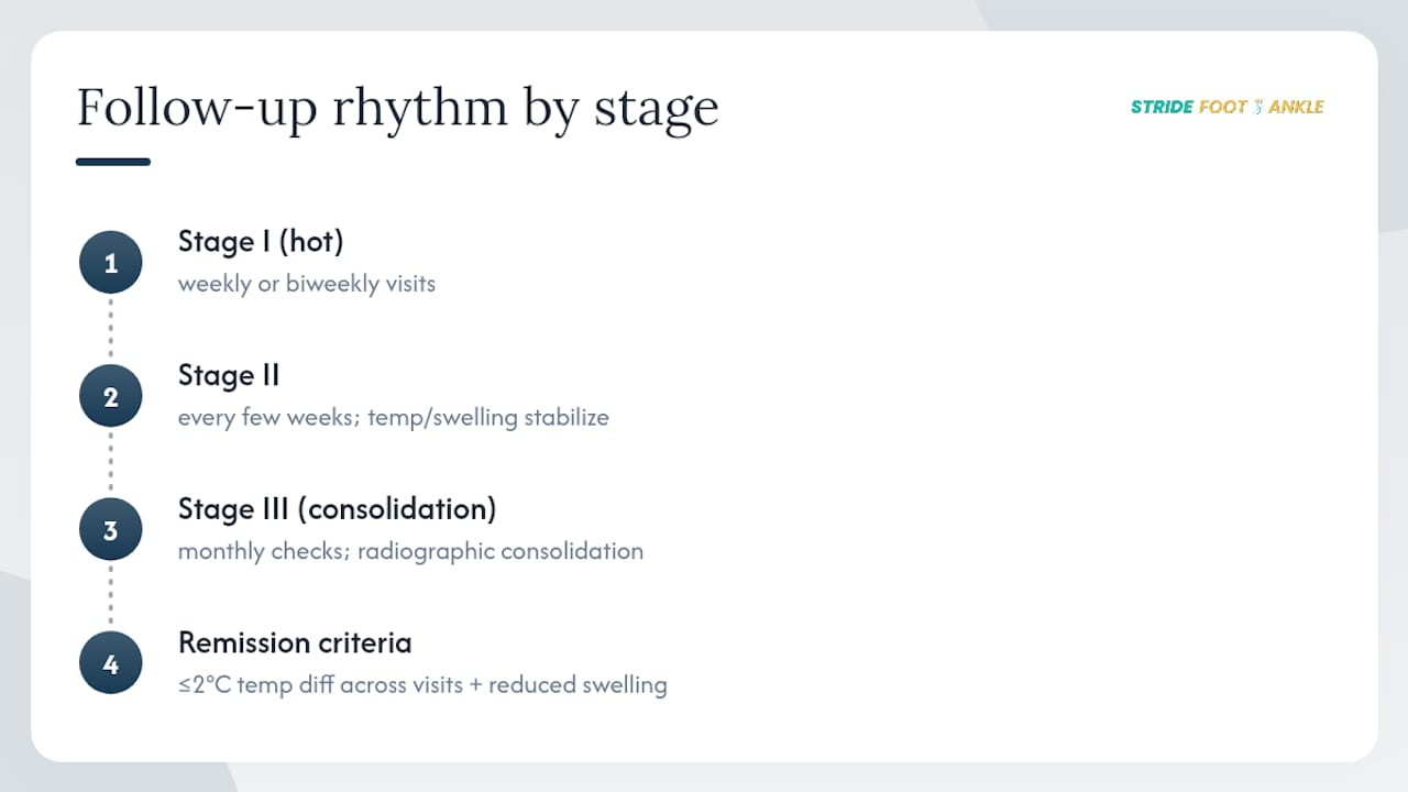
Clinicians don’t rely on how a patient feels to judge progress, since neuropathy removes the pain signal that would normally flag ongoing damage. Instead, remission is defined by two measurable markers: a skin temperature difference of 2°C or less between the affected and unaffected foot, confirmed across consecutive visits, plus visibly reduced swelling. Some clinics use infrared thermometers for this; others rely on careful touch comparison during exam. Either way, one good reading isn’t enough. Guidelines call for that temperature stability to hold across repeat visits before easing up on immobilization, because a single normal reading can be a fluke.
Imaging supports this process rather than replacing it. Plain X-rays track whether bone fragments are consolidating into a stable shape. MRI or nuclear bone scans get added when the diagnosis is unclear, particularly when clinicians need to distinguish active Charcot from a bone infection, since the two can look strikingly similar on a standard X-ray.
A typical follow-up rhythm looks like weekly or biweekly visits during the hot Stage I phase, stretching to every few weeks as temperature and swelling stabilize through Stage II, then monthly checks during Stage III consolidation. Each milestone, temperature normalization, swelling resolution, radiographic consolidation, has to be met before stepping down to a less restrictive device. Rushing that sequence is one of the more common reasons a foot that seemed to be healing suddenly flares back up.
What Long-Term Foot Protection Prevents Relapse?
Once a Charcot foot reaches consolidation, it never fully returns to normal bone strength, so permanent protective footwear becomes a lifelong requirement rather than a temporary phase. The goal shifts from healing an active process to preventing a new one from starting in a foot that remains structurally more fragile than it looks.
Protective options scale with deformity severity. Mild cases often do well in custom, multidensity insoles and extra-depth shoes with a rocker sole that reduces pressure on the forefoot with each step. More significant deformities, especially rocker-bottom feet from midfoot collapse, typically need a CROW boot, a rigid, custom-molded device that functions almost like a removable cast for daily wear. Our overview of bracing options for foot and ankle conditions walks through how these devices are fitted and adjusted over time.
Daily home monitoring matters just as much as the footwear itself:
- Check both feet every evening for redness, warmth, blisters, or new swelling, using a mirror if needed to see the sole.
- Compare temperature between feet by touch, or with an infrared thermometer if your clinic recommends one.
- Inspect shoes and orthoses for wear patterns that suggest a new pressure point developing.
- Never walk barefoot, even at home, since neuropathy means you won’t feel a stepped-on object.
Beyond home checks, plan on clinic visits every three to six months indefinitely, more often if anything changes. That routine surveillance, paired with the ulcer prevention strategies that apply to any neuropathic foot, is what keeps a consolidated Charcot foot from reactivating years down the line.
What Are the Warning Signs of a Charcot Emergency?
Certain changes mean same-day evaluation, not a wait-and-see approach. Treat any of the following as urgent:
- A wound that’s draining, foul-smelling, or rapidly enlarging. This suggests infection, not simple irritation.
- Fever, chills, or feeling generally unwell alongside foot swelling, which points toward a systemic infection risk.
- Redness spreading beyond the immediate area, especially if it’s tracking up the leg.
- Sudden new or worsening pain, which is notable precisely because neuropathy usually blunts pain, so a spike is significant.
- Any change in color or warmth suggesting compromised circulation, such as a foot turning pale, blue, or unusually cold.
When infection is suspected, the treatment priority flips. Instead of continuing routine offloading, clinicians typically pursue a deep tissue culture or bone biopsy, start targeted or empiric antibiotics, and bring in surgical or wound care specialists urgently for debridement. Infection combined with Charcot collapse substantially raises amputation risk if source control is delayed, which is why these situations don’t wait for a scheduled follow-up.
What to Expect From a Clinical Evaluation for Charcot Foot
Stride Foot & Ankle approaches suspected Charcot cases the way the evidence says they should be handled: urgent immobilization first, imaging and specialist coordination in parallel, and a surgical opinion only when conservative bracing genuinely can’t stabilize the foot. Bring a list of your medications, recent blood sugar readings if you track them, and any prior foot X-rays to your first visit.
That visit typically includes a physical exam comparing both feet, temperature and swelling assessment, and imaging orders to establish a baseline. If surgery is ever on the table, we evaluate operative candidacy around glucose control, circulation, and infection status before anything else.
The Gap Between Guideline Recommendations and Real-World Adherence
The IWGDF’s push for nonremovable offloading exists precisely because removable devices fail so often in practice, not because they’re clinically inferior on paper. A patient who can take off a boot will, especially once the foot stops hurting, and neuropathy means it stops hurting long before it’s actually healed. If you take one thing from a Charcot diagnosis, let it be this: insist on the nonremovable option, plan your work and family life around several months of limited mobility, and treat lifelong protective footwear as non-negotiable, not optional once things “feel fine.”
— Ramil
Get Evaluated for Charcot Foot in Las Vegas
If you’re noticing unexplained swelling, warmth, or redness in one foot and you live with diabetic neuropathy, waiting for it to resolve on its own is the one thing that turns a manageable inflammatory episode into permanent deformity. Stride Foot & Ankle offers same-track evaluation for suspected Charcot cases, meaning Dr. Nahad Wassel can order imaging, apply offloading, and coordinate with your endocrinologist or vascular specialist without you bouncing between separate offices first.
Your first visit covers a full foot and ankle exam, imaging to establish baseline bone position, and an immediate immobilization plan if active Charcot is suspected. From there, care is coordinated across general foot and ankle services, covering everything from casting and bracing through surgical evaluation if conservative treatment doesn’t hold. If you’ve noticed swelling that won’t quit or a foot that looks different than it did last month, schedule an evaluation now rather than waiting for a follow-up appointment that’s still weeks away.
This article is general information, not a substitute for advice from a qualified doctor. Consult a qualified healthcare professional about your own circumstances before acting on anything here.
Sources
- Guidelines on the diagnosis and treatment of active Charcot neuro-osteoarthropathy in persons with diabetes mellitus (IWGDF 2023)
- Charcot Neuropathic Osteoarthropathy – StatPearls – NCBI Bookshelf
FAQ
Can I Walk With a Charcot Foot?
Weight-bearing during the active, hot phase is generally restricted or avoided entirely because pressure accelerates bone destruction. Your clinician will guide you through a gradual return to protected weight-bearing as swelling and temperature stabilize.
How Long Does It Take for a Charcot Foot to Heal?
Most people reach clinical remission in four to six months of consistent offloading, though the complete process, from acute swelling through consolidation and transition to protective footwear, can extend to about a year.
What Should You Avoid With a Charcot Foot?
Avoid walking barefoot, skipping offloading device wear time even when the foot feels fine, and using devices like compression boots without checking contraindications first, since certain compression modalities aren’t appropriate for compromised circulation or active inflammation.
What Happens if Charcot Foot Goes Untreated?
Untreated Charcot typically progresses to severe midfoot or ankle collapse, a rocker-bottom foot deformity, chronic ulceration, and a substantially higher risk of infection and amputation. Early nonremovable offloading is what prevents that outcome, which is why suspicion alone, before imaging confirms anything, is enough to start treatment.
Recent Comments