

A foot stress fracture is a hairline crack in a bone caused by repeated impact rather than a single injury. It most often shows up in the metatarsals, navicular, calcaneus, or sesamoids. If you have point tenderness and pain that builds with activity but eases with rest, stop high-impact exercise and get evaluated. Waiting can turn a manageable injury into a complete fracture.
TL;DR:
- Most foot stress fractures occur in the metatarsals, navicular, calcaneus, or sesamoids, with the location influencing treatment complexity and recurrence risk.
- Early-stage stress fractures may not appear on initial X-rays, making MRI the preferred imaging tool for diagnosis during the first few weeks.
- Conservative treatment typically heals low-risk fractures within 6 to 8 weeks, but high-risk sites like the navicular may require surgery or immobilization.
- Preventing recurrence involves gradual training increases, addressing biomechanics, and improving bone health through nutrition and systemic risk factor management.
- Accurate diagnosis and tailored management are crucial for avoiding complications like nonunion or refracture, especially in high-risk foot bones.
Table of Contents
- How Foot Stress Fractures Develop and Where They Strike
- Symptoms and Risk Factors: When Foot Pain Is a Stress Fracture
- Diagnosis and Imaging: Why the First X-ray Often Looks Normal
- Treatment and Management by Risk Group and Fracture Site
- Recovery Timeline and Getting Back to Full Activity
- Preventing Foot Stress Fractures: Bone Health and Training Fixes
- What to Expect During a Stress Fracture Evaluation
- Get Evaluated for Persistent Foot Pain
- Sources
- FAQ
How Foot Stress Fractures Develop and Where They Strike
Bone is living tissue that constantly breaks down and rebuilds itself in response to load. A stress fracture happens when repetitive submaximal stress outpaces the bone’s ability to repair itself, and microscopic damage accumulates faster than remodeling can keep up. Add fatigued muscles into the mix, and the problem compounds. Tired calf and foot muscles stop absorbing shock the way they should, shifting more force directly onto bone.
This is why the classic stress fracture patient isn’t someone who fell or twisted an ankle. It’s a runner who jumped their mileage up too fast, a dancer doing back-to-back rehearsals, or a new military recruit grinding through boot camp. Sports-medicine literature consistently points to these three groups as the highest-incidence populations, largely because all three combine high repetitive loading with limited recovery time between sessions.
Certain bones in the foot take the brunt of this more than others.
- Metatarsals, especially the second and third, are the most common site, particularly among runners and dancers whose forefoot absorbs repeated push-off force.
- Navicular, a small bone on top of the midfoot, carries a disproportionate share of load during running and sprinting and has a notoriously poor blood supply.
- Calcaneus (heel bone) fractures show up frequently in new recruits and walkers who suddenly increase distance.
- Sesamoids, the two tiny bones under the big toe joint, are vulnerable in dancers and sprinters who load the forefoot repeatedly.
Site matters enormously for how these injuries behave. A calcaneal stress fracture and a navicular stress fracture might feel similar on day one, but they carry very different risks and require different treatment timelines, which is the subject of the role imaging plays in podiatry diagnosis and the risk-stratification section further down this article. Metatarsal stress fractures, in particular, have a notably high recurrence rate when the underlying risk factors, like training errors or biomechanical issues, aren’t addressed, according to a StatPearls review of stress reactions and fractures.
Symptoms and Risk Factors: When Foot Pain Is a Stress Fracture
Foot stress fracture symptoms follow a recognizable pattern: pain that starts during or after activity, tenderness at a specific point you can press on with one finger, and mild swelling. Pain that improves with rest but returns whenever you resume the activity that caused it is the hallmark that separates a stress fracture from ordinary soreness or generalized overuse pain.
Generalized overuse pain tends to be diffuse. You can’t point to one spot. It often loosens up as you warm up, then fades by evening. A stress fracture behaves the opposite way: it gets worse the longer you’re on it, and unlike a muscle strain, it rarely responds to stretching or foam rolling. The American Academy of Orthopaedic Surgeons’ OrthoInfo resource notes that pain worsening with weight-bearing activity, paired with focal tenderness, is one of the clearest clinical signs distinguishing a true stress fracture from soft-tissue overuse.
Some symptoms should push you toward urgent evaluation rather than a wait-and-see approach:
- Pain present at rest or that wakes you up at night
- Pain that keeps intensifying over several days despite reduced activity
- Numbness, tingling, or a cold, pale foot (possible nerve or circulation involvement)
- Visible deformity or inability to bear any weight at all
Pro Tip: Try the “one-finger test.” If you can point to a single spot on the bone that reproduces sharp pain when pressed, that’s a stronger signal of a stress fracture than pain that’s spread across a wider area. Bring that detail to your appointment. It genuinely speeds up the exam.
Several risk factors make some people far more likely to develop these injuries than others. A sudden jump in training volume or intensity, the classic “too much, too soon” pattern, is the single most common trigger. Foot structure matters too: people with high, rigid (cavus) arches transmit more shock straight into bone, while those with excessive supination load the outer foot unevenly.
Systemic factors deserve just as much attention as biomechanics. Female athletes dealing with the Female Athlete Triad or Relative Energy Deficiency in Sport (RED-S) face substantially elevated fracture risk because low energy availability disrupts hormone levels that bones depend on for normal remodeling. Low bone mineral density, whether from RED-S, aging, or other metabolic causes, has the same effect. Smoking impairs bone healing and blood flow, and worn-out or poorly cushioned footwear removes one of your body’s few natural shock absorbers. Any stress fracture that occurs without an obvious training trigger deserves a closer look at these underlying contributors rather than treatment of the bone alone.
Diagnosis and Imaging: Why the First X-ray Often Looks Normal
Diagnosing a stress fracture in the foot starts with a physical exam, not a scan. A clinician checks for point tenderness by pressing directly on the suspected bone, and may use a squeeze test or ask you to hop on the affected foot to see if it reproduces your pain. These maneuvers are quick, inexpensive, and often the first real clue.
Imaging is where things get more nuanced, and where a lot of patients get confused. A plain X-ray is usually the first study ordered, but it frequently comes back clean in the early weeks of a stress fracture. That’s not a diagnostic failure. It’s how the injury behaves. Microscopic bone damage doesn’t show up on a radiograph until enough bone remodeling has occurred to create a visible line, which can take two to three weeks or longer.
Because of that gap, Mayo Clinic’s guidance on stress fracture diagnosis points clinicians toward MRI when a fracture is suspected but the X-ray is negative. MRI picks up the earliest stage of injury, called a stress reaction, by detecting bone marrow edema before an actual crack has formed. That earlier detection window matters: catching a stress reaction before it progresses to a full fracture can shave weeks off recovery.
Different imaging tools serve different purposes:
- X-ray: fast and cheap, but often negative in the first two to three weeks; better for confirming an established fracture later in the process.
- MRI: the most sensitive option for early stress reactions and marrow edema; the current evidence review on foot stress fractures identifies it as the preferred study when early detection changes management.
- CT scan: less sensitive for early marrow changes but excellent for visualizing cortical bone detail, which matters most at sites like the navicular or proximal fifth metatarsal when surgery is being considered.
- Bone scan: highly sensitive but nonspecific; occasionally used when MRI isn’t available or when multiple sites need to be screened at once.
For fractures that happen with minimal trauma, recur, or occur in a patient without an obvious training-related cause, additional workup, such as a DEXA scan to check bone density or blood work to screen for vitamin D deficiency or metabolic bone disease, is often appropriate. This ties directly back to the RED-S and bone health discussion above; a fracture with no clear mechanical explanation is a signal to look deeper, not just to brace the foot and move on.
Treatment and Management by Risk Group and Fracture Site
Foot stress fracture treatment isn’t one-size-fits-all. It depends heavily on where the fracture sits, because some locations heal reliably with rest while others carry a real risk of nonunion, meaning the bone never fully knits back together, and may need surgery from the start.
Low-risk vs. high-risk sites
Clinicians divide foot stress fractures into two broad risk categories, and this distinction drives almost every treatment decision that follows.
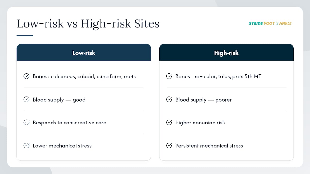
Low-risk sites include the calcaneus, cuboid, and cuneiform bones, along with most metatarsal shaft fractures. These generally have good blood supply and respond well to conservative activity modification alone.
High-risk sites include the navicular, talus, and the proximal fifth metatarsal (the base of the pinky toe bone, sometimes called a Jones fracture). According to the current evidence review on foot stress fracture management, these locations have notably higher nonunion rates and often require a lower threshold for surgical discussion, partly because of poorer local blood supply and partly because of the mechanical forces that continue to stress the site even during protected weight-bearing.
Conservative care for low-risk fractures
For most low-risk stress fractures, treatment follows a fairly consistent path:
- Activity modification: cutting out the impact activity that caused the injury, at minimum for several weeks
- A rigid-soled shoe or walking boot to limit motion at the fracture site and reduce painful flexion
- Protected or modified weight-bearing, sometimes with crutches initially if pain is significant
- Over-the-counter analgesia for pain control (nonsteroidal anti-inflammatories are used cautiously and only under guidance, since some evidence suggests they may slow bone healing)
- Alternative conditioning, like stationary cycling or pool running, to maintain cardiovascular fitness without loading the injured bone
Statistic Callout: Conservative management typically heals low-risk foot stress fractures in about 6 to 8 weeks, according to Cleveland Clinic’s overview of stress fracture treatment. Returning to impact activity before that window closes is one of the most common reasons these injuries drag on or recur.
When immobilization or surgery enters the picture
High-risk fractures often need a stricter approach from day one, sometimes including a non-weight-bearing cast rather than a removable boot, precisely because these patients can’t be trusted (through no fault of their own; it’s just harder to control unconscious loading) to stay off the foot enough in a boot alone. Surgical fixation, typically with a screw or plate to compress and stabilize the fracture, is considered when:
- The fracture shows signs of nonunion on repeat imaging
- The site is high-risk and the patient needs a faster, more predictable return (common in competitive athletes)
- Conservative treatment has already failed once
- The fracture pattern itself is unstable
Evidence for adjunctive therapies, like bone stimulators, shockwave therapy, or ultrasound, remains limited and inconsistent. They’re occasionally used for slow-healing fractures, but they shouldn’t be viewed as a substitute for offloading the bone properly in the first place, a point echoed in the StatPearls clinical review of stress reactions and fractures.
The most common complications when treatment goes wrong, or when patients return to activity too early, are nonunion and refracture. Both are largely preventable with proper risk stratification and patience during the healing window. For readers weighing whether their current care plan fits a conservative or more aggressive path, nonsurgical options for tendon and ligament injuries covers a related decision-making framework worth understanding.
Recovery Timeline and Getting Back to Full Activity
Most low-risk foot stress fractures heal enough for a gradual return to activity within 6 to 8 weeks, though high-risk sites or surgically treated fractures often take considerably longer, sometimes three to four months before full sport clearance. The exact timeline depends on fracture location, how quickly it was caught, and how strictly activity was modified in the early weeks.
Returning too early is the single biggest threat to a full recovery. The safest approach follows a few consistent principles.
- Confirm a pain-free baseline first. You should be able to walk normally, without a limp or lingering ache, before adding any impact back in.
- Increase load gradually. Start with walking, progress to a run/walk pattern, then build toward continuous running or your sport-specific movements over several weeks, not days.
- Match your pain-free duration to your rehab duration. If you were pain-free at rest for two weeks before starting to test activity, plan on a comparably gradual, multi-week progression back into full training rather than jumping straight to prior volume.
- Address strength and gait deficits. Weeks of protected weight-bearing weaken calf muscles and can subtly change how you walk. Skipping strength work sets you up for a second injury.
- Get clinical clearance before high-impact return. This might include a repeat exam, or repeat imaging in select cases, particularly for high-risk sites or athletes returning to competitive-level loading.
Rehabilitation isn’t just about waiting out the calendar. Progressive loading exercises, calf and intrinsic foot strengthening, and gait retraining all play a role in making sure the bone that healed doesn’t get re-injured the moment you’re back on the field. Cadence adjustments (often increasing step rate slightly to reduce per-step impact) and footwear changes can meaningfully lower reinjury risk for runners specifically. The guide to improving foot mobility walks through several of these exercises in more detail, and if this isn’t your first foot or ankle injury, the recovery guide for previous foot and ankle injuries addresses how prior injuries change your risk calculus going forward.
Pro Tip: Return-to-sport decisions are rarely just “wait until it doesn’t hurt.” Clinicians often use a rule of matching your pain-free period to an equal period of graded activity before full clearance. If you were symptom-free for three weeks before testing a light jog, expect a similarly paced, multi-week build back to full training, not an overnight jump.
Preventing Foot Stress Fractures: Bone Health and Training Fixes
Preventing a stress fracture, or a repeat one, comes down to fixing whatever combination of training load, bone health, and biomechanics caused the first one. Treating the bone without addressing the underlying cause is why recurrence rates for these injuries stay stubbornly high.

Anyone who develops a stress fracture without an obvious training spike, or who has already had more than one, should be screened for RED-S or low energy availability. This is especially relevant for competitive runners, dancers, and other athletes in sports that emphasize leanness. When fractures happen with minimal load or keep recurring, referral for bone density testing (a DEXA scan) and bloodwork checking vitamin D and other markers is a reasonable next step, not an overreaction. The research on Female Athlete Triad and RED-S makes clear that treating the fracture without treating the underlying energy deficit just sets up the next one.
On the training side, prevention is mostly about pacing:
- Increase mileage or training volume gradually, generally no more than about 10% per week, rather than making sudden jumps after time off or a new season starting
- Build in cross-training days (cycling, swimming, pool running) to reduce cumulative impact while maintaining fitness
- Track training load, not just distance, factoring in surface, intensity, and terrain changes
- Replace worn running shoes on a regular cycle rather than waiting until they visibly fall apart
- Consider orthotics if you have a notably high arch, excessive supination, or other biomechanical patterns that concentrate load unevenly
The guide to high arched feet breaks down when a cavus foot type warrants custom orthotics versus off-the-shelf support. Nutrition and general bone health deserve attention too. The partner resource on foundational bone health habits covers calcium, vitamin D, and lifestyle factors that support long-term bone strength beyond just the current injury. As the StatPearls review puts it in essence, no single fix, not orthotics alone, not rest alone, not nutrition alone, prevents recurrence as well as addressing all three together.
What to Expect During a Stress Fracture Evaluation
When a patient comes in with suspected foot pain from repetitive stress, the visit starts with a focused history: when did the pain start, what changed in training or daily activity, and where exactly does it hurt. That history often narrows down the likely bone before any exam maneuver even happens.
The physical exam follows next: targeted palpation for point tenderness, functional tests like hopping or a squeeze test, and a look at overall foot alignment and footwear wear patterns. Imaging decisions are made pragmatically. If the story and exam strongly suggest a stress fracture and the location is high-risk, moving straight to MRI often makes more sense than waiting on a likely-negative X-ray first.
The treatment philosophy leans conservative whenever the fracture site allows it: activity modification, protective footwear, and a clear rehab progression cover most cases without surgery. Surgical evaluation gets introduced early and directly for the high-risk sites where waiting carries real cost, rather than being treated as a last resort after conservative care has already failed.
Patients coming in for a first visit should bring a list of recent training changes, any prior foot injuries, and their regular footwear if possible. Most evaluations, including exam and imaging coordination, can be completed within one to two visits, depending on what imaging is needed and how quickly results come back.
— Ramil
Get Evaluated for Persistent Foot Pain
Stride Foot & Ankle gives you a faster, clearer path to a diagnosis than trying to guess your way through rest, ice, and hoping the pain resolves on its own. Because early X-rays frequently miss stress fractures, guesswork at home often means weeks of lost training time and a higher chance of the injury progressing to a full break. Dr. Nahad Wassel evaluates foot pain in person, coordinates the right imaging the first time, and builds a treatment plan around your specific fracture site and activity goals.

If you’re dealing with activity-related foot pain that hasn’t resolved with a few days of rest, or point tenderness you can press directly on, it’s worth getting evaluated rather than pushing through it. Services available include:
- Diagnostic imaging coordination (X-ray, MRI, or CT referral based on suspected fracture site)
- Conservative management with boots, activity modification, and rehab guidance
- Custom orthotics for biomechanical risk factors like high arches or excessive supination
- Surgical care for high-risk fractures, including navicular and proximal fifth metatarsal injuries
Visit Stride Foot & Ankle’s foot and ankle care page to request an appointment and get a clear plan for your recovery.
This article is general information, not a substitute for advice from a qualified doctor. Consult a qualified healthcare professional about your own circumstances before acting on anything here.
Sources
- Stress fractures – Mayo Clinic (Diagnosis & treatment)
- Stress Reaction and Fractures – StatPearls
- Stress Fractures: Symptoms, Causes & Treatment — Cleveland Clinic
FAQ
Can you get a stress fracture from walking?
Yes. Sudden increases in walking distance, especially in worn footwear or on hard surfaces, can cause a stress fracture, and this pattern is common in new military recruits and people starting aggressive walking programs.
How do you heal a stress fracture in the foot?
Most low-risk foot stress fractures heal with activity modification, a protective boot or rigid-soled shoe, and protected weight-bearing for about 6 to 8 weeks; high-risk sites may need immobilization or surgery.
Can you still walk with a stress fracture in your foot?
Many people can walk with a low-risk stress fracture, though it typically causes pain that worsens with continued weight-bearing; high-risk sites like the navicular often require significantly reduced or no weight-bearing.
What are the symptoms of an ankle or foot stress fracture?
The main symptoms are activity-related pain, a specific tender spot you can press on, and mild swelling, with pain that improves during rest and returns with repeated loading.
How do doctors diagnose a stress fracture in the foot?
Diagnosis starts with a physical exam for point tenderness, followed by imaging; because early X-rays often miss these fractures, MRI is frequently used to detect bone marrow edema before a visible crack appears.
Recent Comments