

Bluish or purple discoloration of the feet, called cyanosis, signals that tissues are receiving less oxygenated blood than they need. Most cases in adults are peripheral and caused by cold exposure, vasospasm, or poor circulation, but some are medical emergencies. Check for these red flags right now and act accordingly:
Seek emergency care immediately (call 911 or go to the ED) if you notice any of these:
- Blue or purple discoloration of the tongue, inner lips, or gums
- Sudden breathlessness or chest pain alongside blue feet
- One or two toes turning painful, blue, or black without an obvious cold trigger
- Discoloration that does not blanch (turn white) when you press on it and is spreading rapidly
- Signs of skin breakdown, open sores, or a foul odor from the affected area
If none of those red flags are present: move to a warm room, remove tight footwear, and gently rewarm the foot. If color does not return promptly, call your podiatrist or an urgent care clinic the same day.
Key Takeaways
Cyanotic feet require prompt triage: a pink tongue with symmetric cold-triggered discoloration is usually benign, but a blue tongue, sudden painful toe discoloration, or non-blanching skin demands emergency care without delay.
| Point | Details |
|---|---|
| Central vs. peripheral distinction | A blue tongue signals central cyanosis and requires emergency evaluation; a pink tongue with blue feet points to a peripheral cause. |
| Top red flags for emergency care | Sudden painful blue or black toe, blue tongue or lips, breathlessness, or chest pain all require calling 911 immediately. |
| Blue toe syndrome risk | Asymmetric, painful toe discoloration is often embolic and can progress to necrosis without prompt vascular assessment. |
| Prevention priorities | Smoking cessation, blood glucose control, and protective footwear are the highest-yield steps to reduce recurrence. |
| Stridefootankle in Las Vegas | Stride Foot & Ankle offers same-visit vascular exam, ABI, wound care, and urgent vascular referral for patients with cyanotic feet. |
Table of Contents
- What cyanosis is and why the central vs. peripheral distinction matters
- How cyanotic feet and toes typically look
- Common causes of bluish discoloration of the feet
- How clinicians evaluate cyanotic feet: exam and tests to expect
- Immediate care and treatment options for cyanotic feet
- When you need emergency care right now
- Conditions that look like cyanotic feet but are not
- How cyanotic feet present differently across patient groups
- What to expect for recovery and how to reduce your risk
- What to expect at a podiatry visit for blue or discolored feet
- What I see most often, and what it means for you
- Stride Foot & Ankle is ready to evaluate your feet in Las Vegas
- Sources
- FAQ
What cyanosis is and why the central vs. peripheral distinction matters
Cyanosis is the bluish or purplish discoloration of skin or mucous membranes caused by an elevated concentration of deoxygenated hemoglobin in the blood. Clinicians split it into two categories because the distinction directly determines how urgent the situation is and which organ system to investigate first.
Central cyanosis means the blood leaving the heart is itself poorly oxygenated. It affects the entire body, including the tongue and inner lips. A blue tongue is the single most reliable bedside sign of central cyanosis and points to a cardiac or pulmonary cause requiring urgent evaluation. According to StatPearls on central and peripheral cyanosis, a pink tongue in a patient with blue extremities strongly suggests peripheral rather than central cyanosis, which is a clinically meaningful distinction that changes the entire workup.
Peripheral cyanosis means the blood leaving the heart is adequately oxygenated, but the extremities are not receiving enough of it. The tongue stays pink. Causes range from harmless cold-induced vasospasm to serious arterial occlusion. StatPearls on cyanosis notes that peripheral cyanosis can result from cold exposure, vasomotor changes, peripheral artery disease, or reduced cardiac output.
Pro Tip: Check your tongue in a mirror or have someone look at it. Press a fingernail bed firmly for five seconds and release — color should return within two seconds. Slow capillary refill combined with a blue tongue means central cyanosis until proven otherwise. Call 911.
How cyanotic feet and toes typically look
Cyanotic feet present as a blue, purple, or dusky gray discoloration of the skin, most visible on the toes, nail beds, and the ball of the foot. The color can range from a faint lavender tinge in mild cold-induced cases to a deep violaceous or near-black hue in severe ischemia.
Key visual clues to note and report to your clinician:
- Color tone: Blue-purple in vasospasm; violaceous or mottled in embolic disease; dusky gray or black in necrosis
- Symmetry: Both feet equally affected suggests a systemic or cold-related cause; one toe or one foot points toward a local vascular problem
- Temperature: Cold to the touch favors vasospasm or arterial occlusion; warm with swelling suggests venous congestion
- Pain: Painless and symmetric is more reassuring; sudden, severe pain in a single toe is a red flag
- Blanching: Pressing on cyanotic skin that turns white and then refills is a better sign than skin that stays blue under pressure
- Associated changes: Livedo reticularis (a lacy, net-like purple pattern), petechiae (pinpoint red or purple spots), or ulceration alongside blue discoloration all raise concern for embolic or vasculitic disease
When describing your findings to a clinician, note when the discoloration started, whether it came on suddenly or gradually, whether pulses are palpable at the ankle and top of the foot, and any associated symptoms such as leg swelling, recent trauma, or a new medication.
Common causes of bluish discoloration of the feet
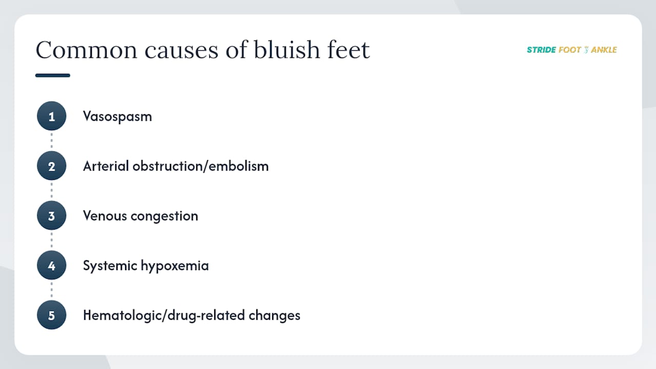
The most likely causes fall into five groups: vasospasm, arterial obstruction or embolism, venous congestion, systemic hypoxemia, and hematologic or drug-related changes. Understanding which group fits your situation helps clinicians order the right tests quickly.
Vasospasm
Raynaud’s phenomenon causes a classic triphasic color change: white (pallor from arterial spasm), then blue (cyanosis from deoxygenation), then red (reactive hyperemia on rewarming). It affects multiple digits symmetrically and is triggered by cold or emotional stress. Primary Raynaud’s is benign; secondary Raynaud’s, associated with connective tissue diseases like scleroderma or lupus, requires further workup.
Acrocyanosis is a persistent, symmetric, painless bluish discoloration of the hands and feet triggered by cold. Primary acrocyanosis is benign and needs only cold avoidance. Secondary acrocyanosis can signal autoimmune disease, malignancy, or an eating disorder and warrants targeted lab testing including autoantibodies and inflammatory markers.
Arterial obstruction and embolism
Blue toe syndrome (BTS) is an acute, often painful blue or violaceous discoloration of one or more toes without generalized cyanosis. The most common cause is cholesterol crystal emboli breaking off from atherosclerotic plaques in the aorta or iliac arteries and lodging in small digital vessels. Risk factors include hypertension, smoking, and recent vascular procedures. Peripheral pulses can remain palpable even when toes are critically ischemic, because the occlusion affects small distal vessels rather than major arteries. A systematic diagnostic framework groups BTS etiologies into vascular obstruction, impaired venous outflow, abnormal circulating blood, vasospasm, infection, and drug-induced causes, which guides targeted testing.

Peripheral artery disease (PAD) causes chronic poor circulation in feet from atherosclerotic narrowing of the leg arteries. Patients typically report cramping pain with walking (claudication) and have diminished or absent foot pulses.
Venous congestion
Deep vein thrombosis (DVT) usually presents with swelling, warmth, and pain rather than the cool, pulseless presentation of arterial occlusion. Severe venous congestion can produce a dusky discoloration, but the distinguishing features are the warmth and edema rather than the cold, pale, or pulseless findings of arterial disease.
Systemic hypoxemia
Heart failure, chronic obstructive pulmonary disease (COPD), pulmonary embolism, and congenital heart defects can all reduce the oxygen saturation of blood reaching the extremities. These causes typically produce central cyanosis with tongue involvement alongside the blue feet.
Hematologic and drug-related causes
Methemoglobinemia, caused by certain medications (nitrates in high doses, some anesthetics, dapsone, topical benzocaine) or toxins, produces a characteristic chocolate-brown blood color and a cyanotic appearance that does not improve with supplemental oxygen. Sulfhemoglobinemia behaves similarly. Specific lab testing is required to identify these conditions.
How clinicians evaluate cyanotic feet: exam and tests to expect
The diagnostic goal is to distinguish peripheral from central cyanosis, identify limb-threatening ischemia or an embolic source, and rule out systemic or hematologic causes. A clinician will typically start with a focused history and vascular exam before ordering tests.
The history covers onset (sudden vs. gradual), symmetry, pain, cold triggers, smoking history, recent procedures, medications, and systemic symptoms. The physical exam includes pulse palpation at the femoral, popliteal, dorsalis pedis, and posterior tibial arteries; capillary refill; skin temperature; and inspection of the tongue and mucous membranes.
Diagnostic tests used in evaluation include pulse oximetry, arterial blood gas, ankle-brachial index, duplex ultrasound, and angiography, each providing complementary information.
| Test | What it measures | Abnormal result suggests |
|---|---|---|
| Pulse oximetry | Peripheral oxygen saturation | Below 95% raises concern for systemic hypoxemia |
| Arterial blood gas (ABG) | Blood oxygen, CO2, pH; methemoglobin level | Low PaO2 indicates systemic hypoxia; elevated methemoglobin confirms methemoglobinemia |
| Capillary refill time | Speed of blood return after pressure | Longer than normal suggests reduced perfusion |
| Ankle-brachial index (ABI) | Ratio of ankle to arm systolic pressure | Below 0.9 indicates significant arterial disease |
| Duplex ultrasound | Arterial and venous blood flow | Absent or reversed flow signals occlusion or DVT |
| CT angiography / arteriography | Detailed arterial anatomy | Identifies stenosis, occlusion, or embolic source |
When the workup points to significant PAD, embolic disease, or an unclear source of embolism, referral to vascular surgery is appropriate. A podiatrist is often the first specialist to evaluate cyanotic toes, coordinate ABI testing, order imaging, and initiate wound care while arranging that referral.
Immediate care and treatment options for cyanotic feet
The immediate goals are to restore perfusion, control the source of embolism or thrombosis, and treat hypoxemia or cold-induced vasospasm before tissue damage becomes irreversible.
Immediate first-aid steps:
- Move to a warm environment and gently rewarm cold feet using warm (not hot) water or blankets
- Remove constrictive footwear, socks, or bandages
- Keep the limb at heart level; do not elevate a suspected ischemic limb, as elevation worsens perfusion
- Do not massage a toe suspected of embolic occlusion — it can dislodge further emboli
- Use supplemental oxygen only if you have a prescription or are under medical supervision
Treatment by cause:
- Vasospasm (Raynaud’s, acrocyanosis): Rewarming, avoidance of cold triggers, and vasodilator medications such as calcium channel blockers (e.g., nifedipine) for moderate-to-severe Raynaud’s
- Blue toe syndrome / embolic occlusion: Anticoagulation to prevent further emboli, urgent source workup (echocardiogram, aortic imaging), and possible embolectomy or bypass for limb-threatening ischemia
- PAD: Revascularization through angioplasty, stenting, or surgical bypass; medical management with antiplatelet agents and statins; supervised exercise programs
- DVT / venous congestion: Anticoagulation (typically low-molecular-weight heparin bridging to oral anticoagulants) and graduated compression once arterial disease is excluded
- Ischemic ulcers: Professional wound care including debridement, offloading, and infection management; avoiding self-treatment of foot wounds is critical because home remedies can accelerate tissue loss
Pro Tip: Never apply heat packs directly to an ischemic foot or use unprescribed topical agents on a blue or ulcerated toe. Ischemic tissue has impaired sensation and cannot dissipate heat normally, making burns a real risk. Let a clinician guide local wound care.
When you need emergency care right now
Call 911 or go to the emergency department immediately if blue discoloration of the feet is accompanied by a blue tongue or lips, sudden breathlessness, chest pain, or a single painful toe that turned blue or black without a cold trigger.
The most urgent scenarios are central cyanosis from a cardiac or pulmonary cause, acute limb ischemia from arterial occlusion, and blue toe syndrome with signs of tissue death. Central cyanosis with tongue involvement means the blood leaving the heart is inadequately oxygenated — this is a systemic emergency that requires oxygen, cardiac monitoring, and rapid diagnosis. Acute limb ischemia from a clot or embolus presents with the classic “6 P’s”: pain, pallor, pulselessness, paresthesia, paralysis, and poikilothermia (cold limb). Any combination of these signs in a foot or leg requires emergency vascular evaluation within hours to prevent amputation. A single painful, blue, or black toe that appeared suddenly — especially in a patient with known atherosclerosis or after a vascular procedure — is blue toe syndrome until proven otherwise and should not be dismissed as “poor circulation.”
For non-emergency presentations (symmetric, cold-triggered, painless, color returns with warming), call your podiatrist or a same-day urgent care clinic rather than the ED.
Conditions that look like cyanotic feet but are not
Not every blue or discolored foot is cyanosis. Several conditions mimic the appearance and require different management entirely.
- Bruising and trauma: Ecchymosis from injury is typically brown-yellow at the edges, follows a trauma history, and does not blanch uniformly; it is not associated with pulse changes
- Purpura and vasculitis: Non-blanching red or purple spots (petechiae or purpura) suggest small-vessel vasculitis or a clotting disorder; pressing on the skin does not cause color to disappear, unlike early cyanosis
- Livedo reticularis: A lacy, net-like purple pattern that can accompany cyanosis but also occurs independently in autoimmune disease, antiphospholipid syndrome, or as a benign cold response
- Pigmentary disorders: Post-inflammatory hyperpigmentation, hemosiderin staining from chronic venous disease, or racial/ethnic skin tone variation can produce dark discoloration without any vascular compromise
- Nail discoloration: Fungal infection, trauma, or subungual hematoma can turn nails dark or bluish; nail discoloration causes differ substantially from true cyanosis of the surrounding skin
- Methemoglobinemia and sulfhemoglobinemia: These produce a grayish-blue skin color that does not improve with supplemental oxygen and is confirmed by a co-oximetry panel showing elevated methemoglobin or sulfhemoglobin levels; the blood itself appears chocolate-brown
When a patient’s cyanotic appearance does not respond to oxygen and pulse oximetry reads low despite the patient appearing otherwise stable, methemoglobinemia or sulfhemoglobinemia should move to the top of the differential.
How cyanotic feet present differently across patient groups
Infants, people with diabetes, and patients with chronic cardiopulmonary disease all carry a lower threshold for evaluation because their presentations can be subtler and their complications more severe.
- Newborns: Acrocyanosis of the hands and feet in the first hours of life is normal and resolves as circulation stabilizes. Central cyanosis with a blue tongue in a newborn is never normal and requires immediate pediatric or neonatal evaluation for congenital heart disease. Parents should call their pediatrician or go to the ED without delay if a newborn’s lips or tongue look blue.
- People with diabetes: Diabetic neuropathy masks pain, so ischemia can progress silently to ulceration and infection before the patient notices. Any new blue or dark discoloration of a toe in a diabetic patient warrants same-day podiatric evaluation. Preventing foot ulcers in this population depends on early detection and regular foot surveillance.
- Smokers and patients with known vascular disease: These patients face a heightened risk of blue toe syndrome and Buerger’s disease (thromboangiitis obliterans), a condition almost exclusively linked to tobacco use that causes inflammatory occlusion of small and medium vessels. Any new toe discoloration in an active smoker with vascular disease should be evaluated urgently.
- Older adults with Medicare: Many older patients are unsure whether a podiatry visit is covered. Medicare coverage for podiatry applies to medically necessary foot care, which typically includes evaluation of vascular changes and diabetic foot conditions.
What to expect for recovery and how to reduce your risk
Prognosis depends primarily on the underlying cause, how quickly treatment begins, and whether coexisting conditions like diabetes or heart disease are well controlled.
- Vasospasm (Raynaud’s, primary acrocyanosis): Usually fully reversible with warming and trigger avoidance; long-term prognosis is excellent with appropriate management
- Blue toe syndrome from embolism: Risk of tissue loss and amputation is real without prompt treatment; published case reports document progression from blue toe discoloration to ulceration and necrosis when ischemia goes untreated, while early wound care and smoking cessation have supported healing
- PAD: Revascularization can restore circulation, but outcomes depend on disease severity and patient adherence to risk-factor modification
- Systemic hypoxemia: Prognosis tracks the underlying cardiopulmonary disease; optimizing heart failure or COPD management is the primary lever
Preventive steps that make a measurable difference:
- Stop smoking — it is the single most modifiable risk factor for PAD, blue toe syndrome, and Buerger’s disease
- Control blood glucose, blood pressure, and cholesterol to slow atherosclerosis
- Wear protective, well-fitting footwear, especially if you have diabetes or neuropathy
- Avoid prolonged cold exposure; use insulated footwear and gloves in winter
- Schedule annual vascular or podiatric checks if you have diabetes, PAD, or a history of toe ischemia
What to expect at a podiatry visit for blue or discolored feet
A podiatrist is often the first specialist to evaluate cyanotic feet, and a well-structured visit can distinguish a benign vasospastic cause from a limb-threatening emergency within a single appointment. General foot and ankle care at a podiatric practice covers the full range of vascular, wound, and structural assessments needed for this presentation.
At a typical visit for cyanotic or discolored feet, you can expect:
- A detailed history covering onset, triggers, symmetry, pain, smoking status, medications, and systemic symptoms
- A focused vascular exam: pulse palpation, capillary refill, skin temperature, and mucous membrane inspection
- Bedside ankle-brachial index (ABI) measurement to screen for PAD
- Ordering of duplex ultrasound or referral for CT angiography when indicated
- Wound assessment and a wound care plan if ulceration is present
- Urgent referral to vascular surgery when findings suggest acute limb ischemia or a significant embolic source
Podiatry is the right first call for non-emergency presentations: symmetric cold-triggered discoloration, mild PAD without rest pain, diabetic foot surveillance, and wound care. The emergency department is the right destination when red flags are present (blue tongue, sudden severe pain, signs of tissue death). Understanding why seeing a podiatrist matters for vascular foot concerns can help you make that call with confidence.
What I see most often, and what it means for you
Most patients who come in worried about blue feet have had a cold-triggered vasospastic episode, and the reassurance they need is that their tongue is pink, their pulses are present, and their color returns with warming. That conversation takes about ten minutes and prevents a lot of unnecessary anxiety.
What concerns me more are the patients who wait. A 60-year-old smoker with a single painful blue toe who assumes it will resolve on its own is the patient at real risk of losing that toe. The clinical picture in those cases is unmistakable once you know what to look for: asymmetric, painful, non-blanching, and not triggered by cold. Getting that patient to a vascular team within hours rather than days is what changes the outcome. The difference between a toe that heals and one that does not is often measured in how quickly someone picked up the phone.
Stride Foot & Ankle is ready to evaluate your feet in Las Vegas
If you are in Las Vegas and your feet or toes have turned blue, purple, or dusky, a same-day or next-day evaluation at Stride Foot & Ankle gives you a clear answer fast. Dr. Nahad Wassel provides a complete vascular and podiatric assessment, including pulse exam, ABI measurement, wound evaluation, and imaging coordination, all in one visit. For patients with ischemic ulcers or wounds that need ongoing care, the practice offers dedicated wound care services with a structured treatment plan.

If your presentation includes red flags (blue tongue, chest pain, sudden severe toe pain, or signs of tissue death), go to the emergency department now. For everything else, schedule your evaluation online or call the practice directly. Getting a clear diagnosis early is the step that protects your foot and your long-term mobility.
Sources
The following clinical and patient-facing resources informed this article and are worth reviewing for deeper detail:
- Central and Peripheral Cyanosis
- A review and approach to the diagnosis of blue toe syndrome | Discover Medicine | Springer Nature Link
- Cyanotic Changes of the Toes – PMC – NIH
- Acrocyanosis
FAQ
What does a cyanotic foot look like?
A cyanotic foot appears blue, purple, or dusky gray, most visibly on the toes and nail beds. The color may be symmetric across both feet in cold-related causes or confined to one or two toes in embolic disease.
What causes cyanosis in the legs and feet?
The most common causes include cold-induced vasospasm (Raynaud’s, acrocyanosis), arterial obstruction or cholesterol emboli (blue toe syndrome, PAD), venous congestion from DVT, and systemic hypoxemia from heart or lung disease.
What are blue feet a symptom of?
Blue feet can indicate anything from harmless cold-triggered vasospasm to serious arterial occlusion, heart failure, or pulmonary disease. The urgency depends on whether the tongue is also blue, whether pain is present, and whether the discoloration is symmetric.
Does blue toe syndrome go away on its own?
Blue toe syndrome caused by cholesterol emboli does not reliably resolve without treatment. Without prompt vascular assessment and anticoagulation or intervention, the affected toe can progress to ulceration and necrosis, as documented in published case reports.
This article provides general health information and is not a substitute for professional medical advice. If you are concerned about cyanotic feet or any foot discoloration, consult a qualified healthcare provider or go to the emergency department for red-flag symptoms.
Recommended
- Why You Should Avoid Self-Treating Foot Wounds – Stride Foot & Ankle – Dr. Nahad Wassel
- Foot and Ankle Swelling: Causes, Remedies, and Red Flags – Stride Foot & Ankle – Dr. Nahad Wassel
- Understanding Foot Fungus and When to Seek Help – Stride Foot & Ankle – Dr. Nahad Wassel
- Guide to Common Pediatric Foot Problems: Causes, Signs, Care – Stride Foot & Ankle – Dr. Nahad Wassel
Recent Comments