

Bruising on the bottom of your foot after a sudden, painful event is a clinical red flag for a plantar fascia tear, not just a minor contusion. The key warning signs are a felt or heard “snap,” immediate ecchymosis (black-and-blue discoloration) along the medial arch or heel, a focal tender lump or palpable gap, and difficulty bearing weight. If you have those signs, you need imaging and evaluation, not just rest.
Immediate action plan (first 0–48 hours):
- Protect and offload. Stop the activity. Avoid putting full weight on the foot. Use crutches or a stiff-soled shoe if you must walk.
- Ice and elevate. Apply ice wrapped in a cloth for 15–20 minutes every 2 hours. Keep the foot above heart level when resting.
- Get evaluated. If you cannot bear weight, if bruising is spreading rapidly, or if you felt a snap, contact a podiatrist or go to urgent care the same day.
Pro Tip: Take a photo of the bruising within the first few hours. Ecchymosis fades and migrates, so a timestamped photo gives your clinician the clearest picture of the injury’s extent and location.
Key Takeaways
A plantar fascia bruise with a sudden snap, ecchymosis along the medial arch, and inability to bear weight is a likely rupture requiring prompt imaging and immobilization, not home rest alone.
| Point | Details |
|---|---|
| Rupture vs. fasciitis | Rupture presents with sudden snap, focal bruising, and a palpable gap; fasciitis builds gradually with no ecchymosis. |
| Steroid injection risk | 94% of rupture patients in one cohort had prior steroid injections; discuss injection history with your clinician. |
| Ultrasound first | Ultrasound is the first-line imaging tool; MRI is reserved for surgical planning or complex anatomy. |
| Recovery range | Conservative care returns most patients to activity in 2–26 weeks, with a mean of 9.1 weeks in an athlete cohort. |
| Stride Foot & Ankle | Dr. Nahad Wassel offers same-visit ultrasound, boot prescription, and conservative-first care in Las Vegas. |
Table of Contents
- What a plantar fascia bruise or rupture looks and feels like
- What causes plantar fascia tears and who faces higher risk
- How clinicians confirm a plantar fascia tear: exam and imaging
- Conservative and surgical treatment options
- What recovery commonly looks like
- When to seek urgent care or go to the emergency department
- How Stride Foot & Ankle evaluates and manages suspected plantar fascia tears
- A clinician’s perspective on treating plantar fascia tears
- Stride Foot & Ankle can help you get the right diagnosis and care
- Sources
- FAQ
What a plantar fascia bruise or rupture looks and feels like
A plantar fascia rupture has a distinct symptom pattern that separates it from most other causes of heel pain. Recognizing it early helps you decide how urgently to act.
Core signs of a rupture:
- Sudden, sharp pain at a specific moment during activity or a misstep
- An audible or palpable “snap” at the time of injury
- Immediate swelling and bruising (ecchymosis) along the medial heel or arch
- A focal tender lump or a palpable gap in the fascia
- Reluctance or inability to bear weight without significant pain
Clinical reports consistently document this pattern: sudden pain, a snap, and localized ecchymosis, with ultrasound confirming a gap between torn fibers in the acute phase.
How plantar fasciitis differs. Classic plantar fasciitis builds gradually. The hallmark is sharp morning pain with the first steps that eases after a few minutes of walking. Bruising is uncommon in uncomplicated fasciitis, and tenderness is usually diffuse across the heel rather than pinpointed to a single spot or gap.
Other causes of plantar bruising to consider:
- Heel fat-pad contusion: direct impact to the heel pad, tenderness is broad and central rather than along the arch
- Stress fracture of the calcaneus: deep, aching bone pain that worsens with activity; confirmed by MRI or bone scan
- Vascular or bleeding disorders: spontaneous bruising without a clear mechanism, often bilateral or recurrent
Pro Tip: Press firmly with one finger along the medial arch from the heel toward the ball of the foot. A rupture typically produces one spot of intense, focal tenderness. Write down exactly where it hurts and when the pain started — your clinician will ask.
What causes plantar fascia tears and who faces higher risk
A plantar fascia tear usually results from one of three mechanisms: a single forceful event, chronic degeneration from prior fasciitis, or a direct blow to the arch.
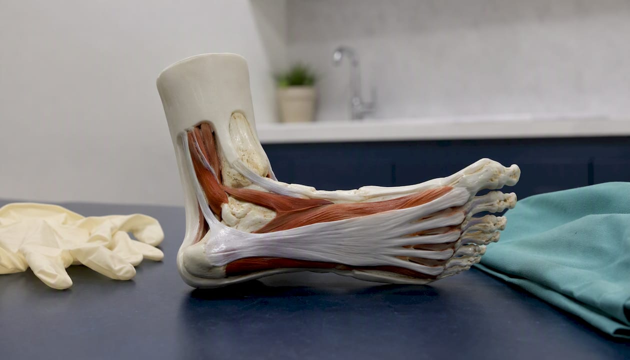
The most common acute mechanism is forceful plantar flexion, such as pushing off hard while sprinting, landing awkwardly from a jump, or catching a foot on a step. Chronic degeneration from longstanding plantar fasciitis weakens the fascia’s collagen structure over time, lowering the force needed to cause a tear. Surgical and sports-medicine case series document both traumatic and degenerative mechanisms in athletes across a range of sports.
The steroid injection connection is the most clinically significant risk factor. A retrospective review of 286 patients with plantar fasciitis found that 33 of 35 patients who sustained a rupture had received prior corticosteroid injections, with a reported odds ratio of approximately 33. That association is striking, even accounting for the retrospective design. A separate systematic review confirms that while corticosteroid injections reduce heel pain over 4–12 weeks, clinicians must weigh those short-term benefits against complications including rupture and fat-pad atrophy.
Additional risk factors:
- Multiple or repeated steroid injections into the same site
- Obesity or high body mass index, which increases tensile load on the fascia
- Sudden spike in training volume or intensity (common in recreational runners)
- Chronic overuse in runners, jumpers, and dancers
- Prior history of plantar fasciitis, which signals pre-existing collagen changes
Statistic callout: In the retrospective cohort, 94% of patients who ruptured their plantar fascia had a history of prior steroid injection — a proportion that underscores why injection frequency and post-injection activity counseling matter.
How clinicians confirm a plantar fascia tear: exam and imaging
Diagnosis starts with the physical exam, and a skilled clinician can often identify a rupture before any imaging is ordered.
Key exam findings:
- Focal tenderness directly over the fascia, often at or just distal to the calcaneal insertion
- A palpable gap or thickened nodule at the tear site
- Visible swelling and ecchymosis along the medial arch
- Pain reproduced by passive dorsiflexion of the toes or the first metatarsophalangeal joint (the windlass test)
- Flattening of the medial arch compared to the uninjured side
Ultrasound vs. MRI: which test and when
A radiology review recommends ultrasound as the first-line modality for plantar fascia disorders, favoring it for its ability to distinguish true fiber interruption from surrounding edema in the acute and subacute phases. MRI becomes the preferred tool when surgical planning requires precise measurement of retraction or when the clinical picture is ambiguous.
| Feature | Diagnostic ultrasound | MRI |
|---|---|---|
| Best use | Acute fiber interruption, local hemorrhage, dynamic exam | Retraction measurement, surgical planning, complex anatomy |
| Speed | Same-day, point-of-care | Scheduled, 30 minutes |
| Cost | Lower | Higher |
| Key advantage | Dynamic assessment, repeatable, no radiation | Superior soft-tissue contrast, full anatomic detail |
| Key limitation | Operator-dependent; limited for deep retraction | Not dynamic; less accessible acutely |
A 2024 case report and review noted that no universally accepted guidelines exist for plantar fascia rupture and recommended early diagnosis with ultrasound and individualized conservative rehabilitation.
Pro Tip: Before your appointment, write down: (1) the exact moment the pain started and what you were doing, (2) whether you felt or heard a snap, (3) a list of any prior steroid injections with approximate dates, and (4) the footwear you were wearing. Bring the shoes if possible. This information cuts the history-taking time in half and helps your clinician order the right imaging immediately.
Conservative and surgical treatment options
Most plantar fascia tears respond well to nonoperative care when managed promptly and correctly. The goal is to protect the healing tissue, restore load tolerance gradually, and address the biomechanical factors that contributed to the injury.
Home care in the first 48–72 hours
- Stop loading the foot. Avoid walking barefoot or in flat, unsupportive footwear.
- Use a stiff-soled shoe or boot. Even before a formal boot is prescribed, a firm-soled athletic shoe reduces fascial strain.
- Ice. 15–20 minutes every 2 hours for the first 48 hours reduces swelling and pain.
- Elevate. Keep the foot above heart level when resting.
- Over-the-counter analgesia. Ibuprofen or naproxen sodium can reduce inflammation and pain for most adults; follow package dosing and avoid exceeding recommended daily limits. Check with your doctor if you have kidney, stomach, or cardiovascular concerns before using NSAIDs.
Typical nonoperative protocol
An athlete cohort treated with boot immobilization and staged weightbearing reported all 18 athletes returned to activity between 2 and 26 weeks, with a mean of 9.1 ± 6.0 weeks, using the following general sequence:
| Phase | Duration (approximate) | Activity level |
|---|---|---|
| Rigid walker boot, non-weightbearing | 2–3 weeks | Crutches; no weight on foot |
| Boot, progressive weightbearing | 2–3 weeks | Gradual weight-bearing in boot |
| Formal physical therapy | Weeks 4–8+ | Stretching, strengthening, gait retraining |
| Return to sport/high-impact activity | Weeks 8–26 (varies) | Based on strength, mechanics, pain |
Timelines vary by tear severity, patient age, and adherence. Nonsurgical options for tendon and ligament injuries such as custom orthotics and bracing are commonly added during the transition out of the boot to support arch mechanics and reduce re-injury risk.
When surgery is considered
Surgery is reserved for a small subset of patients: those with a large, retracted complete tear that fails to bridge conservatively, or high-demand athletes with persistent functional loss after a full nonoperative course. Surgical options typically involve direct repair of the torn ends or, in chronic cases, reconstruction with tendon augmentation. Post-surgical recovery follows a longer timeline than conservative care and requires careful staged rehabilitation.
Statistic callout: A 2022 systematic review covering 155 patients across multiple studies found that most reported cases of plantar fascia rupture were managed conservatively, with generally favorable outcomes.
Pro Tip: Ask your clinician specifically whether your tear is insertional (at the heel bone) or noninsertional (along the mid-arch). The location affects both the boot protocol and your prognosis — insertional tears tend to heal more completely.
What recovery commonly looks like
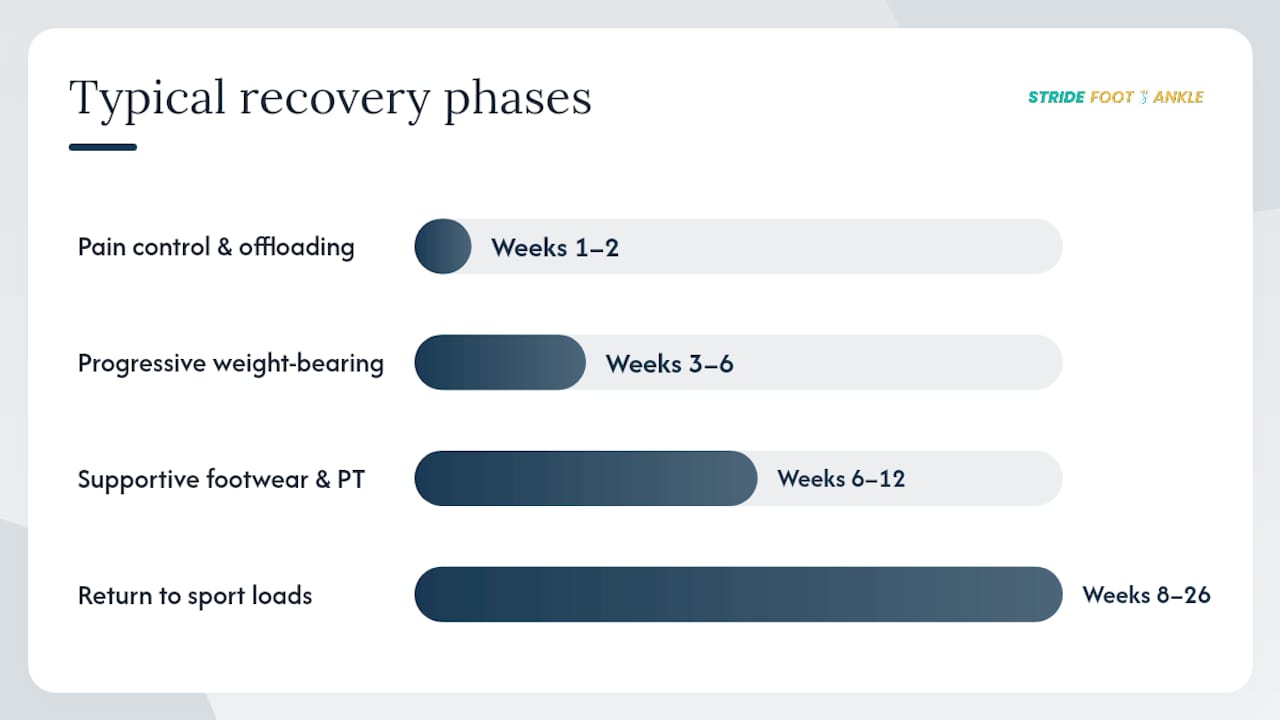
Recovery from a plantar fascia tear follows a predictable arc for most patients, though the pace depends on tear location, completeness, and how quickly immobilization begins.
Typical milestones:
- Weeks 1–2: Pain control improves with offloading; swelling and bruising begin to resolve
- Weeks 3–6: Progressive weight-bearing in boot; walking tolerance increases
- Weeks 6–12: Transition to supportive footwear and orthotics; formal physical therapy addresses strength and gait
- Weeks 8–26: Return to sport-specific loads when strength symmetry and pain-free mechanics are restored
Prognosis differs by tear type. Ultrasound follow-up case series show that partial insertional tears often scar to complete fibrous continuity under conservative care, while noninsertional complete tears may form only partial fibrous bridges. The practical implication: daily-life function usually returns well for both types, but competitive sport performance may be more limited after a complete noninsertional rupture.
Long-term issues to monitor:
- Arch instability or flattening on the injured side
- Altered gait patterns and compensatory loading of the knee or hip
- Lingering difficulty with stairs, hills, or running surfaces
Pro Tip: The two most common reasons recovery stalls are returning to high-impact activity too soon and wearing unsupportive footwear during the rehab phase. Custom orthotics prescribed during recovery help redistribute load across the arch and reduce the risk of re-injury or compensatory problems.
When to seek urgent care or go to the emergency department
Some presentations require same-day evaluation. Do not wait for a scheduled appointment if you have any of the following.
Red flags — contact a podiatrist or go to urgent care immediately:
- Inability to bear any weight on the foot
- Rapidly expanding bruising or swelling within hours of injury
- Severe pain that worsens despite rest, ice, and elevation
- Signs of infection: fever, increasing redness, warmth, or any drainage from the skin
- Progressive numbness, tingling, or loss of circulation (pale or cold foot)
When to go to the emergency department instead of urgent care:
- You cannot bear weight AND have visible deformity (possible fracture or dislocation)
- Signs of vascular compromise: foot is pale, cold, or pulseless
- Rapidly spreading redness with fever (possible cellulitis or necrotizing infection)
Safe interim steps while arranging evaluation:
- Use crutches or a walking boot to keep weight off the foot
- Continue icing (15–20 minutes on, at least 40 minutes off)
- Avoid exceeding recommended NSAID doses while waiting for evaluation
- If you are unsure whether to go to urgent care or the ED, call your podiatrist’s office first; most practices can triage by phone
How Stride Foot & Ankle evaluates and manages suspected plantar fascia tears
At Stride Foot & Ankle in Las Vegas, Dr. Nahad Wassel follows a focused, efficient workflow for suspected plantar fascia injuries. The visit starts with a detailed history: the exact moment of injury, prior steroid injections, activity level, and footwear. That history directly shapes which exam maneuvers and imaging are ordered.
Clinic visit flow:
- Focused physical exam including windlass test, focal palpation for a gap or nodule, and arch mechanics assessment
- Point-of-care ultrasound when the clinical picture suggests fiber interruption or hemorrhage
- MRI referral when retraction measurement or surgical planning is needed
- A treatment plan built around imaging findings and the patient’s functional goals
Common treatments offered:
- Short-term immobilization with a walker boot or controlled ankle motion device, with bracing guidance tailored to the tear type
- Custom or prefabricated orthotics to support arch mechanics during and after recovery
- In-house physical therapy planning or referral for progressive rehabilitation
- Surgical consultation for cases with large retraction or failed conservative care
What to bring to your appointment:
- A written timeline of when symptoms started and what you were doing
- A list of prior steroid injections with approximate dates
- Photos of the bruising taken in the first 24–48 hours
- The footwear you were wearing at the time of injury
- Insurance card and photo ID
Dr. Wassel’s clinical credentials in podiatry include board certification in foot and ankle surgery, with a practice philosophy centered on accurate diagnosis and conservative-first management whenever the clinical picture supports it.
Pro Tip: If you had a prior steroid injection for plantar fasciitis, tell the front desk when you call to book. That history changes the urgency level of your appointment and the imaging ordered at your visit.
A clinician’s perspective on treating plantar fascia tears
Most patients who come in with a plantar fascia bruise are surprised by two things: how dramatic the bruising can look, and how well most tears respond to proper immobilization when caught early. The cases that do best are the ones where the patient protected the foot in the first 48 hours and came in before trying to “walk it off.”
What gives me confidence in conservative recovery is the imaging follow-up data. Partial insertional tears, in particular, tend to scar back to continuity when the fascia is given time and support. The patients who struggle are usually those who delayed immobilization or returned to high-impact activity before strength and mechanics were restored.
My consistent message to patients is this: the timeline is real, and pacing matters more than pushing through. A few extra weeks in a boot is far less costly than a re-rupture or a compensatory knee injury six months later.
Stride Foot & Ankle can help you get the right diagnosis and care
Suspected plantar fascia tears need prompt, accurate evaluation, not a wait-and-see approach. At Stride Foot & Ankle, a single visit with Dr. Nahad Wassel covers the full diagnostic picture: a focused physical exam, point-of-care ultrasound when indicated, and a clear treatment plan that fits your activity level and goals.
Whether you need a walker boot, custom orthotics, a physical therapy plan, or a surgical consult, the clinic offers comprehensive foot and ankle care under one roof in Las Vegas. Patients who are unsure whether their injury warrants a visit can review when and why to see a podiatrist before booking. For those who prefer to start with conservative options, Dr. Wassel’s approach to conservative foot care prioritizes nonoperative management whenever clinically appropriate.
To book an evaluation, visit Stridefootankle or call the Las Vegas office directly. Bring your injury timeline, prior treatment history, and those early bruising photos — the visit will be more productive for it.
This article provides general health information and is not a substitute for professional medical advice. Confirm your diagnosis and treatment plan with a qualified clinician.
Sources
- Plantar Fascia Ruptures in Athletes (cohort report) – PubMed
- Spontaneous rupture of the plantar fascia: case report and rehabilitative approach – Frontiers in Rehabilitation Sciences
FAQ
Can you bruise the plantar fascia?
Yes. A plantar fascia bruise occurs when the fascial fibers tear partially or completely, causing local hemorrhage that appears as ecchymosis (black-and-blue discoloration) along the medial heel or arch. It is a sign of structural injury, not a superficial skin bruise.
What does a ruptured plantar fascia look like?
A rupture typically produces visible bruising along the medial arch or heel, swelling, and sometimes a palpable lump or gap at the tear site. Ultrasound can confirm fiber interruption and local hemorrhage in the acute phase.
How long does a bruised plantar fascia take to heal?
Recovery under conservative care ranges from 2 to 26 weeks depending on tear severity and location, with a mean of approximately 9.1 weeks reported in an athlete cohort treated with boot immobilization and staged physical therapy.
What is stage 4 plantar fasciitis?
Stage 4 plantar fasciitis refers to severe, chronic degeneration of the plantar fascia with significant structural changes, often including partial tearing, calcification, or marked thickening. At this stage, the fascia is at higher risk of complete rupture, and treatment typically requires more aggressive intervention than early-stage fasciitis.
Recent Comments