

A pediatric foot X-ray is a quick radiographic series used to evaluate bone injuries, alignment, and growth-plate concerns in children. Clinicians order it as the first step when a child has foot pain, a suspected fracture, or a gait abnormality. Understanding what these images show, and how to read them, helps both parents and junior clinicians make better decisions faster.
Here is what matters most right now:
- If you are a parent: Keep your child calm and as still as possible during the exam. Bring any prior imaging, a brief description of how the injury happened, and a favorite small toy for distraction.
- If you are a trainee: Confirm the correct views were ordered, correlate findings with the child’s age and ossification stage, and never rely on a single negative X-ray when clinical suspicion remains high.
Resources like Radiopaedia and RadiologyInfo provide solid technical references. For families in Las Vegas who need specialist evaluation, Stridefootankle offers pediatric foot and ankle care guided by board-certified expertise.
Table of Contents
- What are the standard views in pediatric foot X-ray imaging?
- What should parents expect during a pediatric foot X-ray?
- How do you read a pediatric foot X-ray systematically?
- How does normal bone development affect what you see on X-ray?
- What conditions show up on pediatric foot X-rays?
- When is an X-ray not enough, and what comes next?
- Practical tips to get the most out of the imaging visit
- A podiatric surgeon’s perspective on pediatric foot imaging
- Key Takeaways
- The case for careful imaging over convenient reassurance
- Pediatric foot care and specialist evaluation at Stridefootankle
- Useful sources and further reading
- FAQ
What are the standard views in pediatric foot X-ray imaging?
A standard pediatric foot series includes three projections: the dorsoplantar (DP/AP), the lateral, and the medial oblique. Together, these three orthogonal views allow comprehensive evaluation of the phalanges, metatarsals, and tarsal bones in both anatomical and functional positions. The medial oblique is sometimes omitted for straightforward trauma, but it adds real value when metatarsal base or midfoot pathology is suspected.
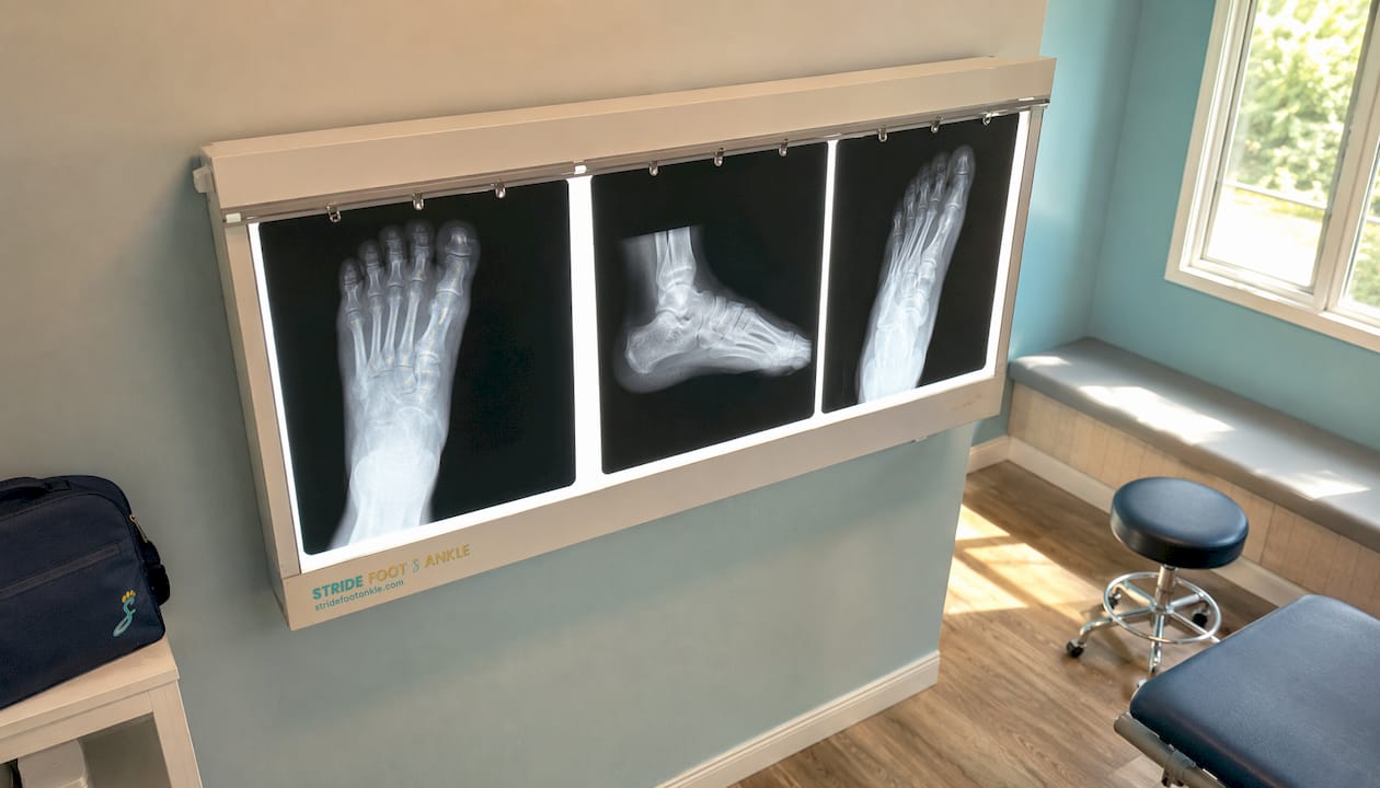
The three standard projections
| View | What it shows | Weight-bearing? |
|---|---|---|
| Dorsoplantar (DP/AP) | Phalanges, metatarsals, midfoot alignment | Preferred weight-bearing |
| Lateral | Hindfoot height, talar declination, calcaneal pitch | Preferred weight-bearing |
| Medial oblique | Metatarsal bases, cuboid, lateral cuneiform | Usually non-weight-bearing |
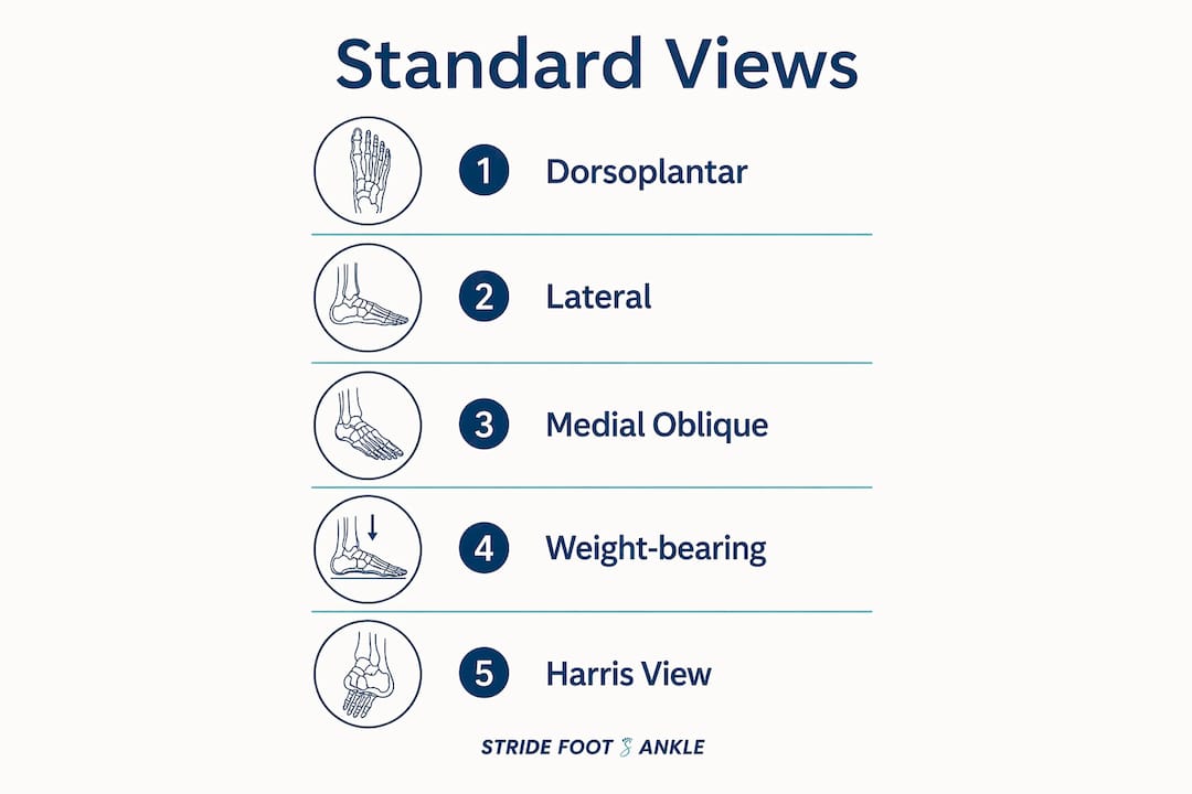
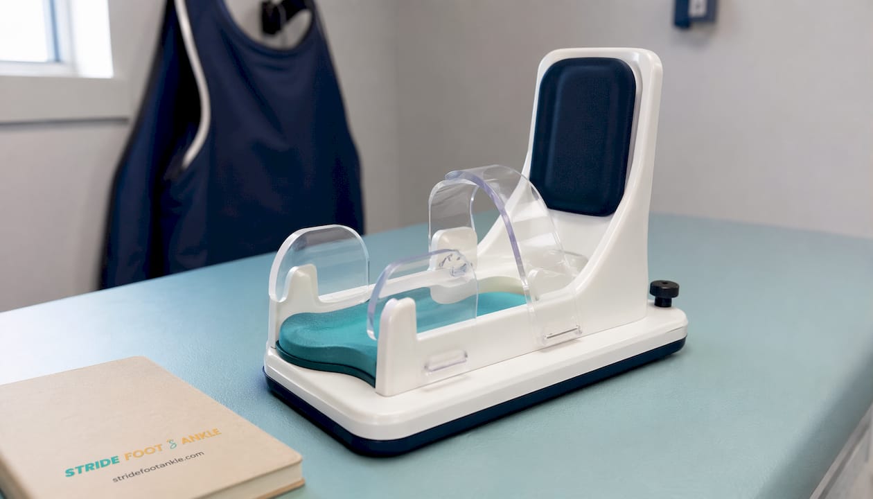
Weight-bearing is the gold standard for alignment assessment. Non-weight-bearing images often fail to show functional malalignment, particularly in flatfoot or equinus deformity. For infants or nonambulatory children, simulated weight-bearing using a rigid board or plantar pressure reproduces functional alignment when true standing views are not possible.
For the DP view, the central beam is typically angled approximately 15° posteriorly toward the heel to reduce bony overlap across the tarsometatarsal joints. The lateral view requires the foot to be placed flat against the cassette with the tibia perpendicular. Exposure times are brief, usually 2–3 seconds per view, which is why stillness matters so much.
A specialized projection worth knowing: the Harris (axial calcaneal) view, with the beam angled 35–45° to the cassette, is used when talocalcaneal coalition is suspected because it better visualizes the posterior subtalar joint and sustentacular facet.
Pro Tip: When ordering a foot series for a child with midfoot pain after a twisting injury, always specify “include medial oblique” on the request. A Lisfranc injury at the second tarsometatarsal joint is easy to miss on DP alone.
What should parents expect during a pediatric foot X-ray?
Pediatric extremity X-rays use very low doses of ionizing radiation. The exam is painless and fast, typically completed within 15 minutes from positioning to image verification. Sedation is rarely needed for foot imaging.
A few things to know before you arrive:
- Remove your child’s shoes, socks, and any metal objects (belt buckles, ankle bracelets) before entering the imaging room.
- Dress your child in comfortable, loose-fitting clothing with no metal hardware near the foot or ankle.
- A guardian can usually stay in the room and will be asked to wear a lead apron.
- Distraction techniques such as swaddling, parental presence, and a favorite toy are the most effective tools for keeping young children still. Radiographers routinely use these instead of sedation.
- Modern protocols focus on fast acquisition and precise positioning to avoid repeat exposures. Whether gonadal shielding is used depends on departmental protocol; some facilities have moved away from routine shielding because it can prolong the exam and increase the chance of motion artifact.
The child must hold still for roughly 2–3 seconds per exposure. That is the single most important thing you can do to help: a calm, still child means fewer repeats and less total radiation.
For children with sensory sensitivities or behavioral challenges, pediatric-focused resources like Child-ology can help families prepare for medical visits in advance.
How do you read a pediatric foot X-ray systematically?
Use a three-step systematic approach: assess alignment first, then evaluate each bone and joint for cortical integrity, then inspect the growth plates and soft tissues. Always correlate with the child’s age and clinical presentation before drawing conclusions.
Step-by-step interpretation checklist
- Verify the study. Confirm the correct side, correct views, and that the images are technically adequate (no rotation, no significant motion artifact).
- Assess alignment. On the lateral view, check the lateral talocalcaneal angle (normal: 35–50°) and the tibiocalcaneal angle (normal: 55–90°). On the DP view, check that the long axis of the talus aligns with the first metatarsal.
- Inspect cortical continuity. Trace each metatarsal shaft and the cortex of each tarsal bone. A subtle cortical step-off or periosteal reaction is often the only sign of a non-displaced fracture.
- Evaluate the physes (growth plates). Look for widening, irregularity, or asymmetry compared to the contralateral side. Physeal widening suggests a Salter-Harris injury even when no fracture line is visible.
- Check the joints. Look for joint space narrowing, effusion, or subluxation, particularly at the tarsometatarsal joints for Lisfranc concerns.
- Assess soft tissues. Soft-tissue swelling localizes injury. Gas in soft tissue suggests infection or an open wound.
Common pitfalls to avoid:
- Ossification centers misread as fractures. Secondary ossification centers are smooth, rounded, and bilateral. Fracture fragments are irregular and do not match a known ossification pattern.
- Non-weight-bearing images masking malalignment. A foot that looks aligned on a supine view may show significant flatfoot or valgus deformity on a standing view.
- Motion artifact mimicking abnormality. A blurred cortex can look like a periosteal reaction. If in doubt, repeat the view with better immobilization.
How does normal bone development affect what you see on X-ray?
Ossification timing matters. Many secondary ossification centers appear and fuse at predictable ages, and they are among the most common fracture mimics in pediatric foot imaging. Knowing the normal sequence prevents unnecessary workup and missed diagnoses.
Ossification centers appear and fuse at various ages during childhood and adolescence, with timing that varies by bone. Awareness of these stages helps to differentiate normal development from pathology.
Common normal variants that cause confusion:
- Os trigonum: A secondary ossification center posterior to the talus, present in a minority of the population. It appears as a smooth, round ossicle and should not be mistaken for an avulsion fracture.
- Calcaneal apophysis: In children aged 7–14, the apophysis normally appears dense and fragmented on X-ray. This is a normal developmental finding, not Sever’s disease on its own. The diagnosis of calcaneal apophysitis requires clinical correlation with posterior heel pain and tenderness.
- Physeal irregularity vs. Salter-Harris fracture: Normal physes can appear irregular, especially at the distal fibula. Asymmetry compared to the other side, combined with point tenderness, is the key differentiator.
For alignment assessment, the lateral talocalcaneal and tibiocalcaneal angles must always be compared to age-appropriate norms. These angles change with development, so a value that is abnormal in a 10-year-old may be normal in a 2-year-old. Pediatric flatfoot evaluation is a good example of where age-normed benchmarks change clinical decisions.
What conditions show up on pediatric foot X-rays?
The most common findings on children’s foot X-ray images range from acute fractures to developmental alignment disorders. Each has a recognizable radiographic signature, and knowing when the X-ray alone is not enough is just as important as recognizing the finding itself.
- Metatarsal shaft fractures: Transverse or oblique cortical break, often with periosteal reaction if subacute. The second and third metatarsals are most commonly involved in stress-type injuries; the fifth is the most common acute fracture site.
- Base of fifth metatarsal avulsion: A cortical step-off or flake at the tuberosity, caused by peroneus brevis traction. The fracture line runs transversely, distinguishing it from the longitudinal apophysis of the fifth metatarsal base in younger children.
- Sever’s disease (calcaneal apophysitis): Fragmentation and sclerosis of the calcaneal apophysis without cortical disruption. Clinical localization to the posterior heel is required for diagnosis; the X-ray appearance alone is not diagnostic.
- Lisfranc injury: Subtle widening of the first and second tarsometatarsal joint space, or a small avulsion flake at the base of the second metatarsal (“fleck sign”). Weight-bearing views are critical here. If clinical instability is present and the X-ray looks normal, MRI is the next step.
- Stress fractures: Periosteal reaction along a metatarsal shaft, sometimes with a faint cortical lucency. Acutely, the X-ray may be completely normal. Repeat imaging at 7–14 days or early MRI is appropriate when clinical suspicion is high.
- Congenital alignment disorders (clubfoot, vertical talus, planovalgus): Assessed on weight-bearing or simulated weight-bearing lateral and AP views using talocalcaneal angle measurements. Clubfoot shows a parallelism of the talus and calcaneus on both views.
Pro Tip: For any child who cannot bear weight after a foot injury but has a normal X-ray, do not discharge with reassurance alone. Occult fractures and physeal injuries are common pediatric foot injuries that X-rays miss acutely. A repeat film at 7–14 days or an early MRI is the safer path.
When is an X-ray not enough, and what comes next?
X-rays are first-line for bone injury and alignment, but a normal radiograph does not exclude soft-tissue injury or complex growth-plate damage. MRI is more sensitive for occult fractures, physeal cartilage injuries, and soft-tissue pathology. CT adds detail for complex bony anatomy and pre-operative planning. Ultrasound is useful for superficial foreign bodies and some soft-tissue assessments.
Order advanced imaging or follow-up when:
- Pain persists despite a negative X-ray and the child still cannot bear weight.
- A physeal injury is suspected and growth-plate arrest would be a significant risk.
- Clinical instability suggests Lisfranc disruption that the X-ray does not confirm.
- Osteomyelitis or a soft-tissue abscess is on the differential.
- A congenital deformity requires pre-operative mapping.
Timing guidance: Repeat X-ray at 7–14 days if fracture suspicion persists after an initial negative film. Periosteal new bone or a fracture line that was radiographically occult acutely often becomes visible by then. Order MRI earlier when physeal cartilage injury or soft-tissue damage is the concern, because waiting two weeks risks missing a window for intervention.
For referral decisions, the role of imaging in podiatry extends well beyond the initial film. Conservative management with bracing is appropriate for many fractures and apophysitis cases. Urgent referral to orthopedics or podiatry is warranted for open fractures, neurovascular compromise, unstable Lisfranc injuries, and suspected growth-plate arrest. Elective referral is appropriate for persistent pain, worsening deformity, or any finding that does not fit a clear benign diagnosis.
Practical tips to get the most out of the imaging visit
Good preparation on both sides of the encounter reduces repeat exposures and improves diagnostic yield.
For parents, before and during the exam:
- Dress your child in shorts or loose pants with no metal hardware near the foot.
- Bring any prior X-rays, MRI reports, or imaging CDs, even from another facility.
- Write down a brief injury history: when it happened, how, and what makes it worse.
- Stay in your child’s direct line of sight during the exam. Your presence is the most effective calming tool available.
- Bring a small favorite toy or allow a short video on your phone for distraction during positioning.
- Accept immobilization aids (foam wedges, tape, sandbags) if the radiographer recommends them. They reduce motion and cut the chance of a repeat exposure.
For junior clinicians ordering the study:
- Specify weight-bearing status on the request form. “Weight-bearing AP and lateral” versus “non-weight-bearing” changes what the radiographer sets up and what the radiologist interprets.
- Include a focused clinical question: “Suspected base-of-fifth avulsion after inversion injury” gives the radiologist context that changes their search pattern.
- Request simulated weight-bearing for nonambulatory children when alignment is the clinical concern.
- Note neurovascular status if the mechanism suggests a high-energy injury.
- Indicate the child’s age clearly. Ossification interpretation is age-dependent, and a missing age can lead to a misread.
Expect a formal radiology report within a few hours for routine studies, or sooner if marked urgent. If the child’s clinical status changes before the report arrives, call the radiologist directly for a verbal preliminary read.
A podiatric surgeon’s perspective on pediatric foot imaging
Radiographs are the starting point for almost every pediatric foot evaluation in clinic, but they are a starting point, not a conclusion. At Stridefootankle, Dr. Nahad Wassel, a board-certified foot and ankle surgeon, uses X-ray findings in direct combination with clinical examination to guide every treatment decision.
Two patterns come up repeatedly in practice. First, a child presents with persistent midfoot pain after a sports injury, the initial X-ray is read as normal, and the family is reassured. When the pain does not resolve in two weeks, a repeat film or MRI reveals a stress fracture or a subtle Lisfranc ligament injury that the first image missed. The lesson: a negative X-ray in a child who cannot bear weight is not a green light to stop investigating. Second, a toddler with a limp and no clear trauma history has a foot X-ray that shows a dense, fragmented calcaneal apophysis. The family is told it looks like a fracture. It is not. It is a normal developmental variant, and the actual diagnosis is a toddler’s fracture of the tibia that the foot film did not capture.
Signs that warrant urgent referral to a podiatric surgeon or orthopedist:
- Open fracture or skin breach over a fracture site
- Neurovascular compromise (absent pulses, capillary refill greater than 2 seconds, paresthesias)
- Clinically unstable Lisfranc injury
- Suspected physeal arrest or growth disturbance
- Progressive deformity that worsens over weeks
Pro Tip: When a child’s X-ray shows a finding that does not fit the clinical picture cleanly, seek a second opinion before committing to a diagnosis. Pediatric foot imaging is one of the areas where a specialist’s eye changes management most often.
Dr. Nahad Wassel brings board-certified surgical training and a patient-centered approach to every pediatric case at Stridefootankle in Las Vegas.
Key Takeaways
Pediatric foot X-rays require age-specific interpretation, weight-bearing views for alignment, and clinical correlation to distinguish normal development from injury.
| Point | Details |
|---|---|
| Standard views | DP/AP, lateral, and medial oblique cover the full foot; weight-bearing views are preferred for alignment. |
| Normal ossification | Many centers (navicular at 3–4 years, calcaneal apophysis at 7–10 years) are common fracture mimics. |
| Alignment benchmarks | Lateral talocalcaneal angle and tibiocalcaneal angle are used as normal reference ranges, varying by age and development. |
| When X-ray is not enough | Repeat at 7–14 days for suspected occult fracture; order MRI earlier for physeal or soft-tissue concern. |
| Stridefootankle | Dr. Nahad Wassel at Stridefootankle in Las Vegas provides specialist pediatric foot evaluation when imaging findings need expert clinical correlation. |
The case for careful imaging over convenient reassurance
There is a temptation in busy clinical settings to treat a normal X-ray as a clean bill of health. For pediatric foot imaging, that shortcut carries real risk. A child’s foot is not a small adult foot. The growth plates, the ossification sequence, the alignment norms, and the injury patterns are all different, and the consequences of a missed physeal injury can follow a child for years.
The radiation exposure from a foot X-ray series is genuinely low, and the clinical information it provides is genuinely high. That balance strongly favors imaging when there is reasonable clinical suspicion. What it does not favor is stopping at the X-ray when the child’s symptoms do not match the film. MRI, repeat imaging, and specialist referral are not signs of over-investigation. They are signs of thorough care.
Parents deserve to know that the exam is safe, fast, and worth doing. Trainees deserve to know that the interpretation requires more than pattern recognition. It requires knowing what a normal 8-year-old’s calcaneal apophysis looks like, understanding why a weight-bearing view changes the diagnosis, and having the clinical confidence to say “the X-ray is negative, but we are not done yet.”
Pediatric foot care and specialist evaluation at Stridefootankle
When a child’s foot X-ray raises questions, or when the diagnosis is clear and treatment needs to start, Stridefootankle offers the specialist care Las Vegas families need. Dr. Nahad Wassel evaluates pediatric foot and ankle conditions with the same board-certified expertise he brings to adult cases, combining imaging review with hands-on clinical assessment to reach the right diagnosis.
Whether the concern is a suspected fracture, persistent heel pain, a gait abnormality, or a finding on imaging that needs a second set of eyes, the clinic provides conservative and surgical options tailored to each child’s age, activity level, and growth stage. Families can learn more about the full range of services at Stridefootankle’s general foot and ankle care page, or review how imaging guides clinical decisions at the imaging in podiatry guide. To schedule a pediatric evaluation, contact Stridefootankle directly to request an appointment.
Useful sources and further reading
These resources support the clinical and patient-facing content in this article. Each is worth bookmarking for ongoing reference.
- Imaging of Pediatric Foot Disorders, PMC/NIH: A peer-reviewed overview of foot imaging across pediatric age groups; useful for trainees building systematic interpretation skills.
- Radiopaedia: Foot Series (Pediatric): Concise technical reference for standard projections, positioning, and protocol notes; the go-to for quick technique review.
- Radiopaedia: Pediatric Foot DP View: Detailed view-specific guidance including beam angulation and positioning tips.
- RadiologyInfo: Pediatric X-Ray Exam: Patient-facing explanation of the procedure, safety, and what to expect; share this link with parents before the visit.
- RadiologyKey: Approach to Pediatric Foot: Clinical reference covering alignment angles, ossification variants, and systematic interpretation; recommended for junior clinicians.
- PMC: Radiographic Assessment of Pediatric Foot Alignment: Peer-reviewed review of weight-bearing assessment and alignment benchmarks across age groups.
- Stanford Children’s Health: X-Rays of the Extremities: Clear patient-facing explanation of what extremity X-rays show and how the procedure works.
- KidsHealth: X-Ray Exam: Foot (for Parents): Practical parental guidance on preparation, what to expect, and how to help during the exam.
Always consult the interpreting radiologist and treating clinician for case-specific findings. General references support understanding but do not replace individualized clinical judgment.
FAQ
What does a pediatric foot X-ray show?
A pediatric foot X-ray shows bone structure, alignment, fractures, and growth-plate status across the phalanges, metatarsals, and tarsal bones. It is the first-line tool for evaluating foot pain, suspected fractures, and alignment concerns in children.
Is a foot X-ray safe for my child?
Yes. Pediatric extremity X-rays use very low radiation doses, and each exposure lasts roughly 2–3 seconds. The diagnostic benefit of identifying a fracture or alignment problem far outweighs the minimal radiation risk from a foot series.
Can a normal foot X-ray miss a fracture?
It can. Occult fractures, physeal cartilage injuries, and soft-tissue damage are not always visible on an initial X-ray. If your child still cannot bear weight after a normal film, a repeat X-ray at 7–14 days or an MRI may be needed.
What is the difference between Sever’s disease and a fracture on X-ray?
Sever’s disease (calcaneal apophysitis) shows fragmentation and sclerosis of the calcaneal apophysis without cortical disruption. A fracture shows a cortical break or step-off. Clinical correlation with the location of tenderness is required because the X-ray appearance of a normal apophysis can look alarming.
When should my child see a podiatric specialist after a foot X-ray?
Seek specialist evaluation when the child cannot bear weight, when imaging shows a growth-plate injury, when pain persists beyond two weeks despite a normal X-ray, or when a deformity is worsening. Stridefootankle provides pediatric foot and ankle evaluation in Las Vegas for exactly these situations.
Recommended
- The Purpose of Foot X-Rays: What You Need to Know – Stride Foot & Ankle – Dr. Nahad Wassel
- What is pediatric foot pain? A parents’ guide to causes and care – Stride Foot & Ankle – Dr. Nahad Wassel
- Pediatric Concerns: Top 10 Foot Issues Parents Should Know – Stride Foot & Ankle – Dr. Nahad Wassel
- Pediatric Ingrown Toenails: A Parent’s Complete Guide – Stride Foot & Ankle – Dr. Nahad Wassel
Recent Comments