

Foot X-rays are the first-line imaging tool for diagnosing bone fractures, dislocations, arthritis, infections, and structural deformities in the foot. They provide rapid, non-invasive visualization of bones and joints, making them the go-to test for unexplained foot pain, acute trauma, and ongoing monitoring of bone conditions.
The core diagnostic purposes of a foot X-ray include:
- Detecting fractures and dislocations after injury or trauma
- Identifying arthritis, bone cysts, and infections such as osteomyelitis
- Evaluating structural deformities like bunions and hammertoes
- Assessing bone alignment, particularly with weight-bearing views
- Guiding treatment decisions, from conservative care to surgical planning
- Confirming proper bone healing after a fracture has been set
What is a foot X-ray and how does it work?
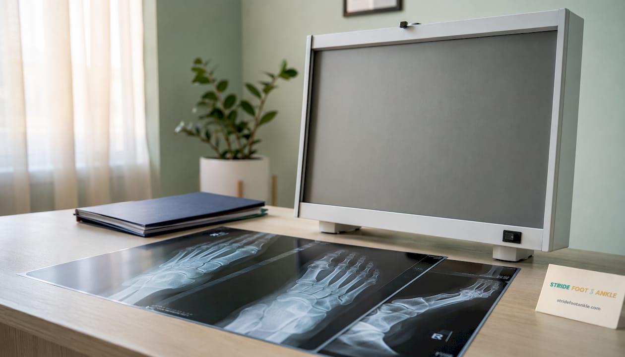
A foot X-ray, also called a foot radiograph or foot series, uses a small, controlled dose of ionizing radiation to produce black-and-white images of the bones and joints in your foot. Dense structures like bone appear white; softer tissues appear in shades of gray. The images go directly into a computer for review, replacing the older film-based process that doctors have used for over a century.
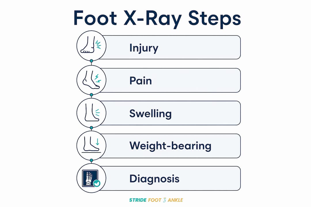
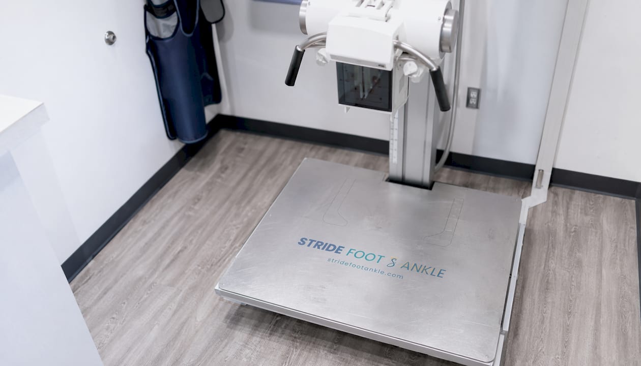
Standard views include the anteroposterior (AP) projection, which looks at the foot from above, and the lateral view from the side. A third oblique view is often added for a more complete picture. For conditions affecting alignment, such as flat foot or Lisfranc injury, weight-bearing views are captured while you stand, placing the bones in their functional position.
Pro Tip: Ask your ordering physician whether you need weight-bearing films. Non-weight-bearing views can miss alignment problems that only appear when you are standing and loading the foot normally.
When is a foot X-ray indicated?
Foot X-rays serve as first-line imaging for nearly all acute foot pain cases. Doctors order them across a wide range of clinical situations:
- Trauma and suspected fractures: Any significant impact, fall, or twisting injury to the foot warrants imaging to rule out a break or dislocation.
- Chronic or unexplained pain: Persistent pain, swelling, or tenderness without a clear cause calls for imaging to identify underlying bone pathology.
- Arthritis evaluation: X-rays reveal joint space narrowing, bone spurs, and erosions that distinguish arthritis from other causes of foot pain.
- Deformity assessment: Bunions, hammertoes, and flat foot are measured and graded radiographically to guide treatment.
- Bone infections: Osteomyelitis produces characteristic changes on X-ray, particularly in diabetic patients.
- Post-treatment monitoring: After a fracture is set or surgery performed, repeat X-rays confirm alignment and healing progress.
Diabetic patients with peripheral neuropathy deserve special mention. Because nerve damage can mask pain, bone pathology may go unnoticed until it is advanced. Radiographic evaluation is advised even without the typical clinical triggers that apply to other patients.
How is a foot X-ray performed?
The entire procedure takes less than 15 minutes and is completely painless. Here is what happens from start to finish:
- You remove your shoes, socks, and any jewelry or metal objects near the foot.
- A radiologic technologist positions your foot on the X-ray platform, adjusting the angle for each required view.
- Depending on the views ordered, you may sit, lie on a table, or stand with your full weight on the foot.
- A lead apron or shield is placed over your body to protect areas not being imaged.
- You hold still for a few seconds while the machine captures each image.
- The technologist reviews the images for clarity before you leave.
Pro Tip: If you are having weight-bearing films taken for a suspected Lisfranc injury, distribute your weight evenly across both feet rather than shifting it. Uneven loading can distort the alignment measurements the radiologist needs.
How should you prepare for a foot X-ray?
Preparation is minimal, but a few steps help the imaging go smoothly:
- Wear comfortable clothing that allows easy access to your foot and ankle.
- Remove all footwear and socks before the exam begins.
- Take off any ankle bracelets, toe rings, or other metal objects near the foot.
- Tell the technologist if you are pregnant or think you might be. Although radiation doses in foot X-rays are low, ionizing radiation carries potential risk to a developing fetus, and protective protocols will be applied.
- No fasting or special diet is required.
What should you expect after the procedure, and is it safe?
Results in an emergency setting are often available immediately. In an outpatient clinic, results typically return within hours to a few days, depending on how the practice is set up. A radiologist reads the images and sends a report to your ordering physician, who then discusses findings with you.
Radiation exposure from a foot X-ray is very low, considered safe for the vast majority of patients. Lead shielding minimizes exposure to surrounding tissues, and the brief duration of the scan limits total dose. Pregnant patients and children may require additional precautions, which the care team will address before imaging begins. There are no known side effects from the procedure itself.
How do X-ray findings guide diagnosis and treatment?
X-ray findings directly shape the clinical path forward. A clearly displaced fracture may lead straight to surgery, while a non-displaced stress fracture points toward rest and protective footwear. Arthritis findings help determine whether conservative management or joint replacement is appropriate. For imaging in podiatry, the X-ray is often the starting point that determines whether additional studies are needed.
Common diagnoses confirmed or identified on foot X-rays include:
- Acute fractures and stress fractures
- Joint dislocations, including Lisfranc disruption
- Osteoarthritis and inflammatory arthritis
- Osteomyelitis (bone infection)
- Bone cysts and benign lesions
- Bunions (hallux valgus) and hammertoe deformities
- Flat foot and other alignment abnormalities
Expert insights on interpreting foot X-rays
Accurate interpretation requires a systematic review of every structure on the film, not just the area of obvious concern. Radiologists and podiatric specialists examine soft tissue swelling, cortical margins, trabecular bone patterns, and joint alignment to avoid missing subtle findings.
Key clinical insight: A systematic approach to reviewing foot radiographs, checking soft tissues, cortical margins, trabecular patterns, and joint alignment, reduces diagnostic errors and improves detection of subtle traumatic findings that might otherwise be missed on a quick scan of the image.
The American College of Radiology recommends radiography as the initial imaging for acute foot trauma, but advises MRI or ultrasound when soft tissue injury is suspected and X-rays are negative. Ligaments and tendons do not appear clearly on X-ray, so a normal film does not rule out a significant soft tissue injury. Understanding X-ray limitations in musculoskeletal care is just as clinically important as knowing what X-rays reveal.
Weight-bearing films carry particular weight in surgical planning. Research published in 2020 found only 45% decision concordance between weight-bearing and non-weight-bearing films for hallux valgus assessment, meaning the choice of imaging position can meaningfully change the surgical recommendation a patient receives.
Foot X-rays at Stridefootankle in Las Vegas
If you are dealing with foot pain, a recent injury, or a condition that has not responded to initial treatment, getting the right imaging is the first step toward real answers.

Stridefootankle, led by Dr. Nahad Wassel in Las Vegas, offers comprehensive foot and ankle care that begins with accurate diagnosis, including on-site imaging and expert interpretation. Rather than guessing at the cause of your pain, Dr. Wassel reviews your imaging findings in the context of your full clinical picture and builds a treatment plan around what the evidence actually shows, whether that means conservative care, orthotics, or surgical correction. Schedule an appointment today to get a clear diagnosis and a path forward.
Key takeaways
Foot X-rays are the standard first-line imaging for acute foot pain and trauma, with weight-bearing views adding critical functional detail that non-weight-bearing films cannot provide.
| Point | Details |
|---|---|
| First-line diagnostic tool | Foot X-rays are the standard initial imaging for nearly all acute foot pain and trauma cases. |
| Weight-bearing views matter | Only 45% decision concordance exists between weight-bearing and non-weight-bearing films for hallux valgus, making view selection clinically significant. |
| Radiation exposure is low | Foot X-ray doses are minimal and considered safe; lead shielding and brief scan duration limit total exposure. |
| Limitations require follow-up | X-rays do not visualize ligaments or tendons well; MRI or ultrasound may be needed when soft tissue injury is suspected. |
| Stridefootankle offers expert care | Dr. Nahad Wassel at Stridefootankle in Las Vegas provides imaging-guided diagnosis and personalized treatment planning for foot and ankle conditions. |
FAQ
How do you tell if you need a foot X-ray?
You likely need a foot X-ray if you have acute trauma, persistent unexplained pain, swelling, or tenderness that does not resolve with basic care. Your doctor uses clinical criteria, including the Ottawa Foot Rules, to determine whether imaging is warranted.
Do you take socks off for a foot X-ray?
Yes. You remove your shoes, socks, and any metal objects near the foot before imaging to prevent artifacts that could obscure the bone detail on the image.
How quickly will I get my foot X-ray results?
Emergency results are often available immediately. In an outpatient setting, results typically return within hours to a few days, depending on the practice and whether a radiologist reads the images on-site.
What foot problems show on X-rays?
Foot X-rays can reveal fractures, dislocations, arthritis, bone infections (osteomyelitis), bone cysts, bunions, hammertoes, and alignment abnormalities such as flat foot and Lisfranc injury.
Recommended
- Why Regular Foot Exams Matter for Your Health – Stride Foot & Ankle – Dr. Nahad Wassel
- The Role of Imaging in Podiatry: A Clinical Guide – Stride Foot & Ankle – Dr. Nahad Wassel
- Why foot care matters: prevent pain, injuries, and complications – Stride Foot & Ankle – Dr. Nahad Wassel
- Why Choose a Foot Specialist for Expert Foot Care – Stride Foot & Ankle – Dr. Nahad Wassel
Recent Comments